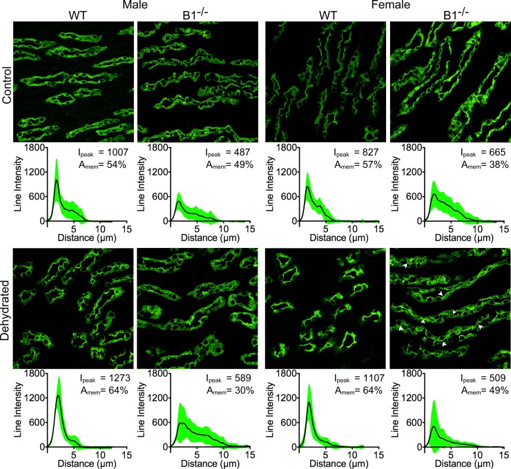
Fig 6. Atp6v1b1-/- animals show a negligible amount of regulation in the base of the inner medulla.
Male WT animals showed a 26% increase in the peak intensity (1st column) and female WT showed a 34% increase in peak intensity (3rd column). In the case of B1-/- males, there was actually a reduction in AQP2 in the membrane region, but the peak intensity increased (2nd column). In the case of female B1-/- animals, there was a considerable reduction in peak intensity (30%), though the protein in the membrane region also increased (29%). In this case, even though the average peak intensity was reduced, there are many cells that had a very clear membrane expression of AQP2 (white arrowheads), indicating a heterogeneous cell regulation of AQP2 (4th column).

