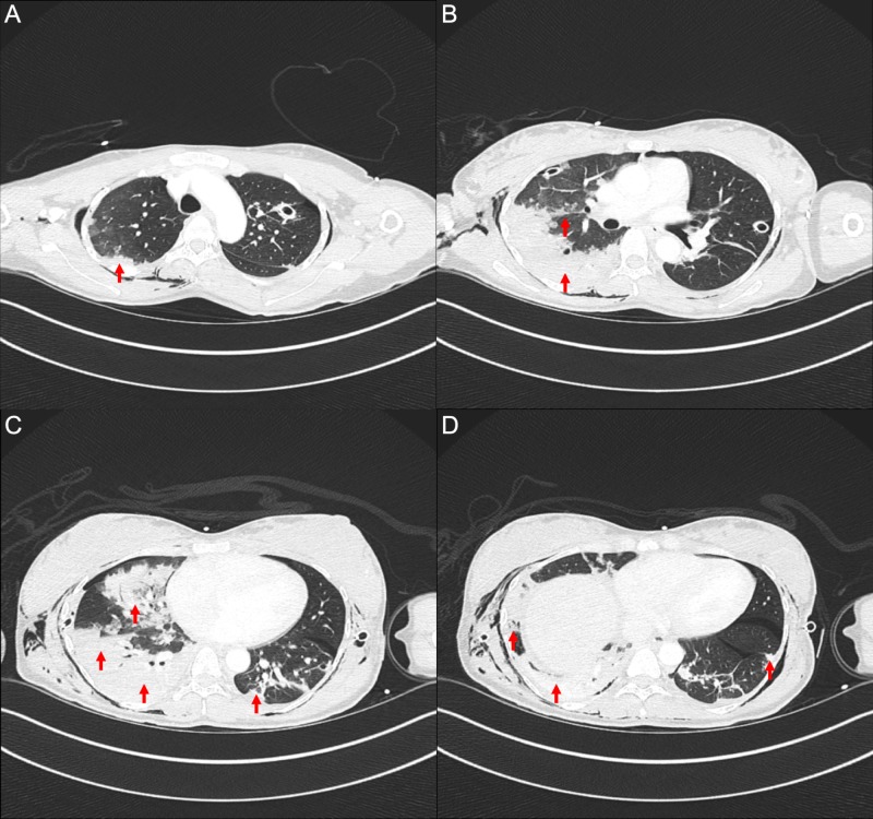
Figure 2. Computed tomography of the chest with iodinated contrast.
Selected axial slices from the apices (A) to lung bases (D) demonstrating extensive, predominantly peripheral consolidations and surrounding ground-glass opacities with a crazy paving pattern present throughout the right lung and left lower lobe.

