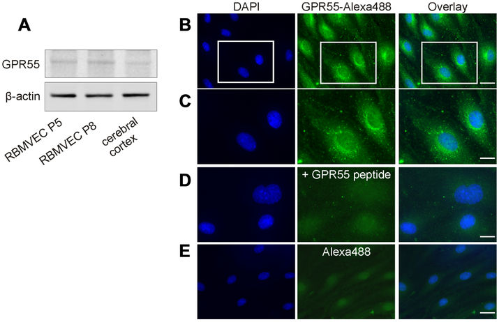
Fig. 1. GPR55 expression and distribution in rat brain microvascular endothelial cells (RBMVEC).
A, Western blot analysis of RBMVEC passage 5 (P5), passage 8 (P8) and rat cerebral cortex indicates the presence of GPR55 at the protein level; β-actin was used as an internal loading control. B. GPR55-like immunoreactivity was found mostly intracellularly; scarce GPR55-like immunoreactivity was seen at the plasma membrane; nuclei are stained with DAPI. C, Higher magnification image of the area outlined in B, illustrating the cellular distribution of GPR55 immunoreactivity. D. Example of control experiments, where GPR55 antibody was incubated with the immunizing peptide; a low basal fluorescence level was detected using Alexa Fluor 488 secondary antibodies. E. Example of control experiments, where GPR55 antibody was omitted, indicating low background fluorescence detected with the secondary antibody Alexa Fluor 488. Scale bar, 20 μm in B and E; 10 μm in C and D.

