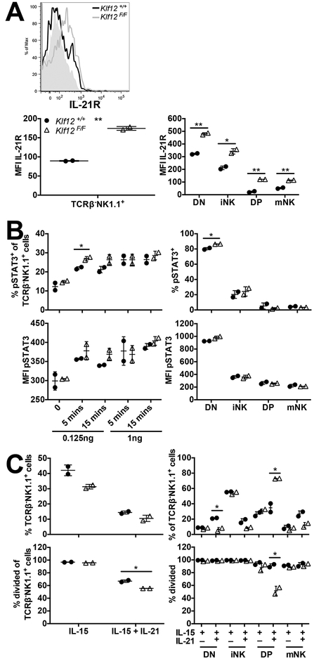
FIGURE 7.
IL-21R expression and signaling are increased in KLF12-deficient NK cells from mixed BM chimeric mice. (A) Representative histogram of IL-21R expression on Klf12+/+ (black line) and Klf12F/F (grey line) splenic TCRβ−NK1.1+ NK cells. The grey-filled histogram is the fluorescence minus one control. MFI of IL-21R expression on splenic TCRβ−NK1.1+ NK cells (left panel) and NK cell developmental subsets (right panel). Data are representative of 2 experiments (n = 2 mice/experiment). (B) Percentage and MFI of pSTAT3 in splenic TCRβ−NK1.1+ NK cells (left panels) upon IL-21 stimulation ex vivo and NK cell developmental subsets (right panels) upon 0.125 ng/ml IL-21 stimulation ex vivo for 15 min. Data are representative of 2 experiments (n = 2 mice/experiment). (C) Percentage of splenic TCRβ−NK1.1+ NK cells (left panels) and NK cell developmental subsets (right panels) after 7 days of in vitro culture in 10 ng/ml IL-15 in the absence or presence of 100 ng/ml IL-21. Data are representative of 2 experiments (n = 2 mice/experiment). *p < 0.05, ** p < 0.005.

