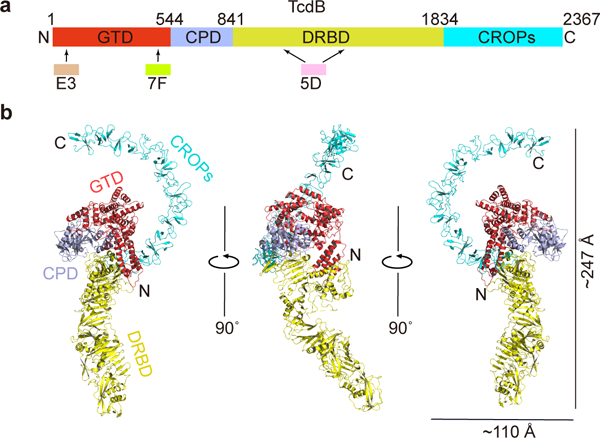
Figure 1.

Overall structure of the full length TcdB holotoxin. (a) A schematic diagram showing the domain organization of TcdB and the approximate VHH-binding regions. GTD, glucosyltransferase domain (red); CPD, cysteine protease domain (light blue); DRBD, delivery and receptor-binding domain (yellow); CROPs, combined repetitive oligopeptides domain (blue). (b) Cartoon representations of TcdB holotoxin. The 3 VHHs that were used to facilitate crystallization were omitted for clarity. TcdB domains are colored as in panel (a).
