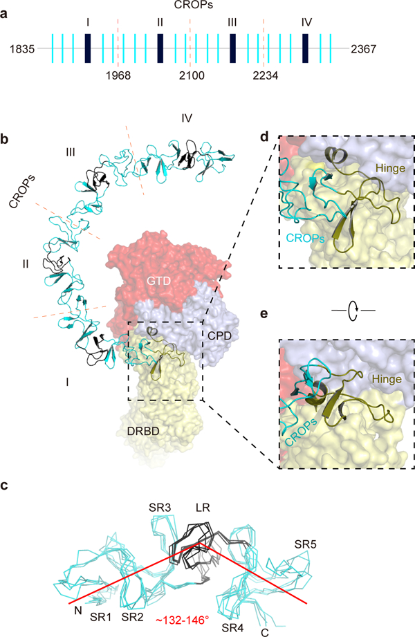
Figure 2.

The unique structure of the CROPs of TcdB. (a) A schematic diagram of the CROPs showing the organization of the short repeats (SRs, thin blue bars) and the long repeats (LRs, thick black bars). The dashed red lines indicate the boundaries of four CROPs units (I–IV). (b) A close-up view into the CROPs while the remainder of TcdB is in a surface representation. (c) Superposition of the 4 CROPs units. The LR in each CROPs unit causes a ~132–146° kink. (d, e) The hinge region (colored olive), which connects the CROPs to the rest of the toxin, is located at the center of TcdB and surrounded by the GTD, the CPD, and the DRBD.
