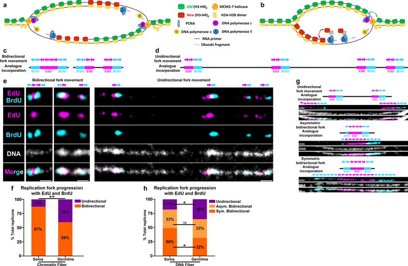
Figure 7: Germline-derived chromatin and DNA fibers show more unidirectional fork progression compared to soma-derived chromatin and DNA fibers.
(a,b) A cartoon showing strand biased histone incorporation at a bidirectional replication fork (a) and a unidirectional replication fork (b). (c,d) Predicted bidirectional fork progression result (c) and unidirectional fork progression result (d). (e) Bidirectional fork progression pattern from somatic cell-derived chromatin fiber. Replicons show early label (EdU) flanked by late label (BrdU) on both sides. Unidirectional fork progression pattern from germline-derived chromatin fiber. Multiple replicons show alternation between early label (EdU) and late label (BrdU) along the chromatin fiber toward the same direction. (f) Quantification of fork progression patterns in somatic cell-derived versus germline-derived chromatin fibers. Testis-derived fibers show a significantly higher incidence of unidirectional fork progression: 42% in testis-derived chromatin fiber (n=54) versus 13% in eye imaginal disc-derived chromatin fiber (n=31) **: P< 0.01, Chi-squared test. (g) Fork progression patterns in DNA fibers: unidirectional fork progression, asymmetric bidirectional fork progression and bidirectional fork progression. (h) Quantification of fork progression patterns in somatic cell-derived versus germline-derived DNA fibers. Germline-derived fibers show a significantly higher incidence of unidirectional fork progression: 35% in germline DNA fiber (n=109), 17% soma DNA fiber (n=48). *: P< 0.05, Chi-squared test. See online Methods for additional statistical information. Scale bar for panels e and g, 1μm

