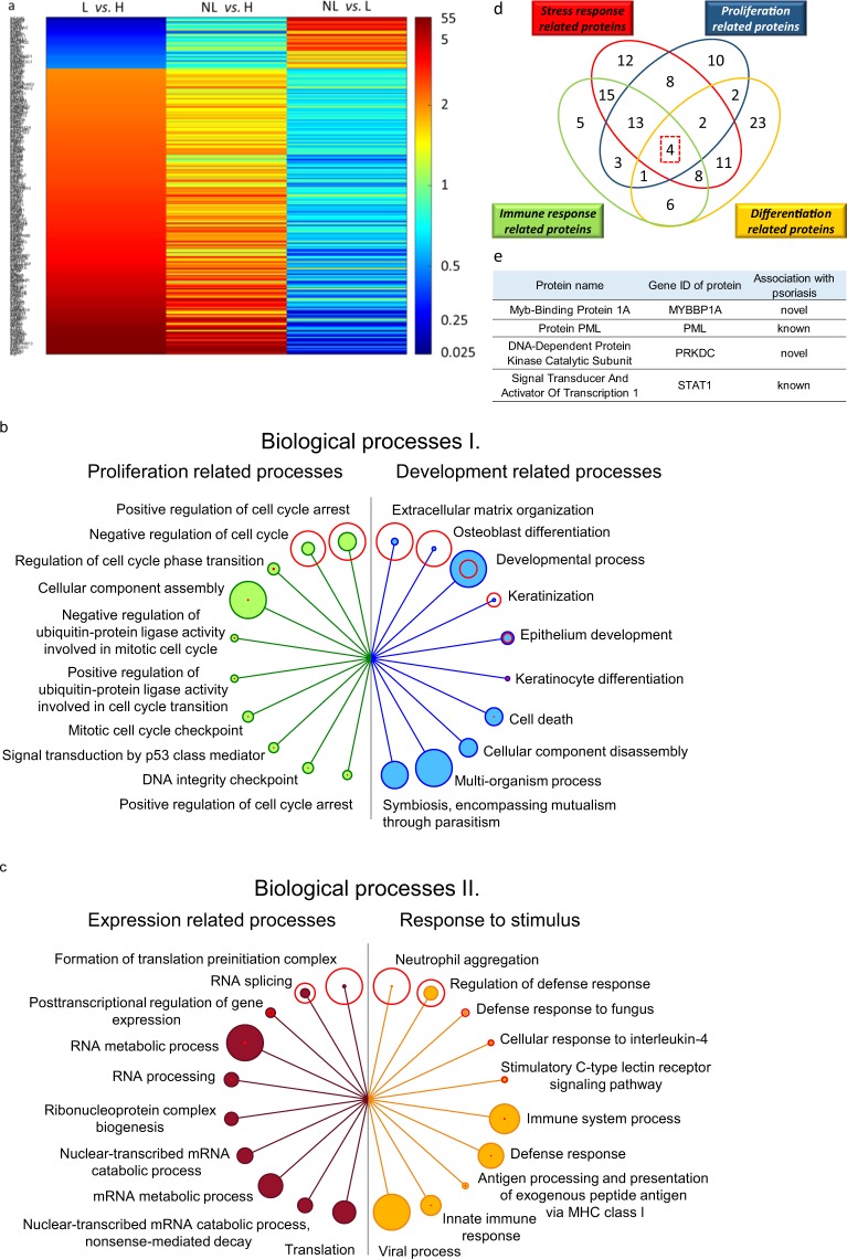
Figure 2.
Characterization of altered protein expression of lesional (L) skin compared to healthy (H) skin. Heatmap of relative expression of proteins differentially expressed in L and H skin (a, left column), and their expression in non-lesional (NL) and L skin (a, middle column) and NL and H skin (a, right column) (a). Biological processes for which proteins were differentially expressed in L and H are listed. The top ten processes are depicted for proliferation (b left, green circles), development (b right, blue circles), expression (c left, filled red circles) and response to stimulus (c right, orange circles). False detection rate (FDR) values are indicated with unfilled red circles around the filled circles for the various biological processes. The size of each circle is proportional to FDR values (unfilled circles) or to the number of proteins (filled circles). Four proteins differentially expressed in H and L skin are believed to participate in all four mechanisms of stress, immune response, proliferation and differentiation (d) and are listed in (e). (*Significant difference in relative protein expression at least by two-fold in L and H comparison).

