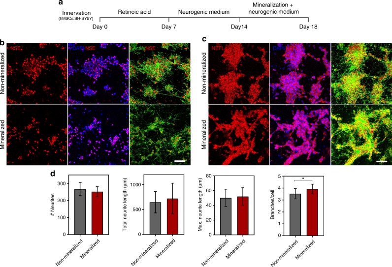
Fig. 5.
Innervation of mineralized cell-laden collagen. a Timeline for the generation of innervated and mineralized collagen constructs. Human SH-SY5Y neuroblastoma cells were co-encapsulated with hMSCs (4:1) in non-mineralized and mineralized hydrogels, after 14 days of differentiation. For the first 7 days, the cells were treated in DMEM/F-12 basal medium supplemented with 1% FBS and 10 µM all-trans retinoic acid (RA), followed by an additional 7 days of culture in Neurobasal-A medium containing 1% (v/v) l-glutamine, 1× B-27 supplement, 50 ng/mL human BDNF, and 10 µM RA. Subsequently, matrix mineralization was triggered by switching the neurobasal medium for the mineralizing medium for another 3 days. The fully differentiated neuronal cells within the mineralized constructs were confirmed by immunostaining with antibodies against Neuron-specific enolase (NSE) and Neurofilament light (NEFL). Cell nuclei were counterstained with DAPI (blue) and cytoskeletal actin was stained with Alexa Fluor 488 phalloidin (green). b, c Representative immunofluorescence images showing the expression of NSE and NEFL in non-mineralized and mineralized constructs. Both the neuronal differentiation markers had similar expression levels in mineralized and non-mineralized groups. Scale bar: 50 µm. d Imaris Filament Tracer module was used to quantify the morphological parameters of the differentiated SHSY-5Y cells in non-mineralized vs. mineralized constructs. Quantification of the number and length of neurites indicate no significant difference between non-mineralized and mineralized groups, whereas the number of branches and branch points was higher in the mineralized groups. Data are represented as Mean ± SD, *p < 0.05, Student’s t-test (N = 4). Source data are provided as a Source Data file

