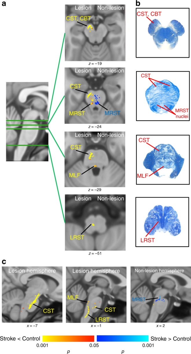
Fig. 1.

Tracts with white matter integrity changes in brainstem in individuals with stroke compared to controls. a Axial view of the tracts with significant decrease (red-yellow) or increase (blue) in white matter integrity in individuals with stroke compared to controls. Corticospinal tract (CST), corticobulbar tract (CBT), medical reticulospinal tract (MRST), lateral reticulospinal tract (LRST), and medial longitudinal fasciculus (MLF) of the lesioned hemisphere showed significant decreased white matter integrity in individuals with stroke compared to controls. Medial reticulospinal tract (MRST) of non-lesioned hemisphere showed significant increased white matter integrity in individuals with stroke. b Brainstem atlas adapted from Gray’s anatomy and Haines’ neuroanatomy with modification for simplicity, showing the location of tracts. c Sagittal view of the tracts with significant decrease (red-yellow) or increase (blue). (Statistical test: two-sample t test with non-parametric 50,000 permutation. Using the threshold-free cluster enhancement with corrected p < 0.05)
