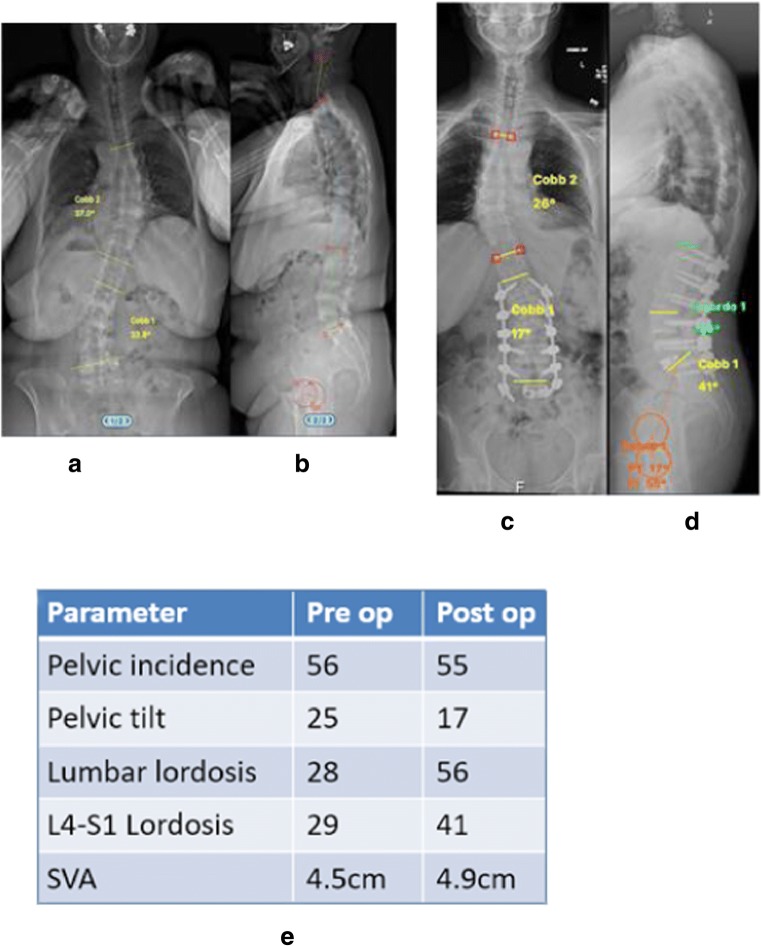
Fig. 2.
Case example of a patient with degenerative lumbar scoliosis who underwent correction with a cMIS approach. a, b Preoperative radiographs. c, d Postoperative radiographs. e Changes in radiographic parameters. Note that a cMIS approach was appropriate for this patient given the small preoperative SVA

