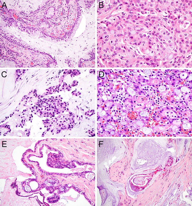
Fig. 3.
Mucin production. Low-grade MEC classically contains prominent goblet cells and cyst formation with abundant luminal mucin (a; × 20); goblet cells can be much more sparse in the oncocytic variant of MEC (b, × 40). CCC frequently demonstrate at least focal mucin production (c, × 40). SC has characteristic luminal secretions that can be both eosinophilic and mucinous (d, × 40). Although salivary duct cysts can demonstrate mucinous metaplasia, the presence of papillary and cribriform elements confirms the diagnosis of MEC (e, × 20). While mucoceles can demonstrate prominent extravasated mucin, floating epithelium with complex architecture favors MEC (d, × 20)

