Fig. 2.
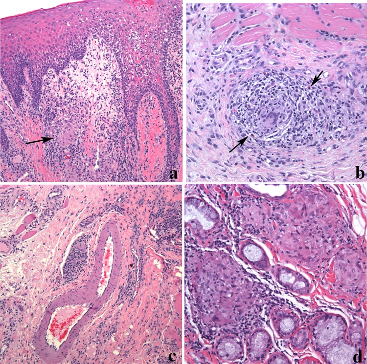
Orofacial granulomatous in a lip biopsy. a Epithelial hyperplasia with inflammatory cell exocytosis overlying edematous connective tissue. Loosely arranged granulomas with histiocytic multinucleated giant cell (arrow) is seen (magnification × 100). b Granuloma composed of a mixture of inflammatory cells including eosinophils (arrow) extending into muscle (magnification × 200). c Granulomas are often present around dilated lymphovascular spaces (magnification × 100). d Granulomas can also extend into minor salivary glands. The granulomas in this image show a nodular collection of epithelioid histiocytes and giant cells surrounded by inflammation (magnification × 200)

