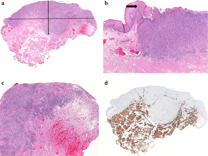
Fig. 1.
a Whole mount image of a lateral tongue squamous cell carcinoma. A horizontal line from adjacent benign epithelium on each side of this tumor would underestimate tumor depth of invasion (DOI). DOI was measured from the surface of the tumor to the deepest point of invasion (arrow). In effect, an “arcuate” line was created to account for the normal contour of the tongue. b Left side of this tumor as seen in 1a, with adjacent benign epithelium. Even though the tumor is not exophytic, the epithelium is drawn upwards, meeting an ulcer. The raised epithelium is a more appropriate choice as the origin for a “horizon” line (arrow). In this case, the line chosen was arcuate across the tumor surface rather than straight across to the opposite side of the tumor. c Right side of this tumor as seen in 1a. Tumor focally undermines adjacent benign epithelium. Choice of anything other than an arcuate line would be quite subjective and would underestimate DOI. d Desmin-stained slide of the same tumor. Skeletal muscle is drawn close to the surface of the mid-portion of the tumor, further justifying the method of DOI measurement described above

