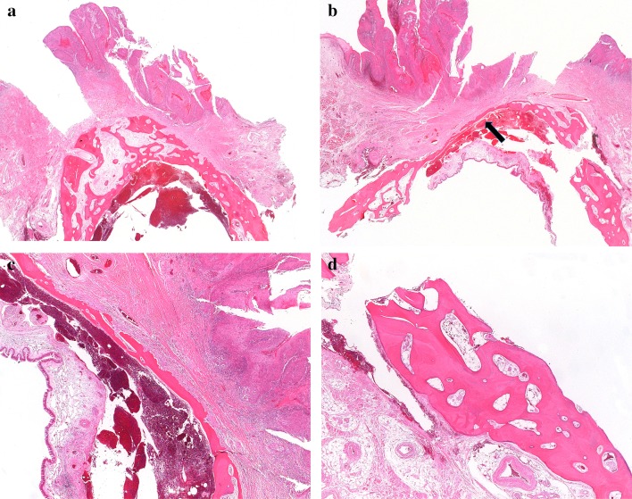
Fig. 3.
a An exophytic maxillary tumor with erosion of the cortical bone of the edentulous alveolar ridge. No bone invasion is identified at this location. b Same tumor at a different location. The leading edge of the tumor has eroded most of the bone, leaving only a thin layer of cortical bone under the sinus mucosa (arrow). Only a small amount of cancellous bone is visible, on the lower left. c The tumor has destroyed most of the bone, with a leading edge of fibroinflammatory tissue and no direct contact between tumor and bone. This was interpreted as bone invasion (category pT4a), but as only minimal cancellous bone is identified nearby, and there is no direct invasion of tumor into the medullary space, that diagnosis is debatable. d Bone of the maxilla from lower right of the field in 3b. In this edentulous patient, the bone is essentially cortical type

