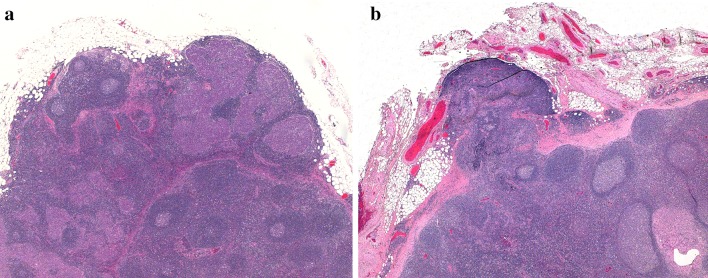
Fig. 5.
a Lymph node with an incomplete capsule and metastatic carcinoma. Although a subcapsular sinus is seen focally (upper left), in other areas the lymphoid tissue merges with perinodal fat. b A lymph node with a capsular defect and protruding lymphoid tissue. Metastatic carcinoma is seen on the lower right, confined to the node

