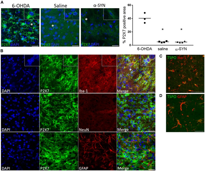
FIGURE 4.

P2X7 protein expression is co-localized with Iba-1 microglial immunoreactivity following 6-OHDA-induced neuroinflammation. (A) A visibly lower P2X7-positive area is shown at the site of injection in the striatum of saline-injected rats at day 14 and the substantia nigra of α-SYN-rats at day 28 post-injection, as compared to the 6-OHDA model. The insets show a magnified image of the cells marked with a star. Scale bar: 25 μm. ∗p < 0.05 Mann–Whitney U test. (B) Representative details of the striatal 6-OHDA lesion core, where increased [11C]JNJ-717 binding was detected at day 14. P2X7 immunoreactivity is co-localized with Iba-1-positive cells of microglia/macrophage lineage, whereas the staining pattern does not coincide with GFAP or NeuN, astrocytic or neuronal markers, respectively. Scale bar: 25 μm. (C,D) A TSPO-positive signal is shown both in microglia and astrocytes. Scale bar: 50 (C) and 35 μm (D).
