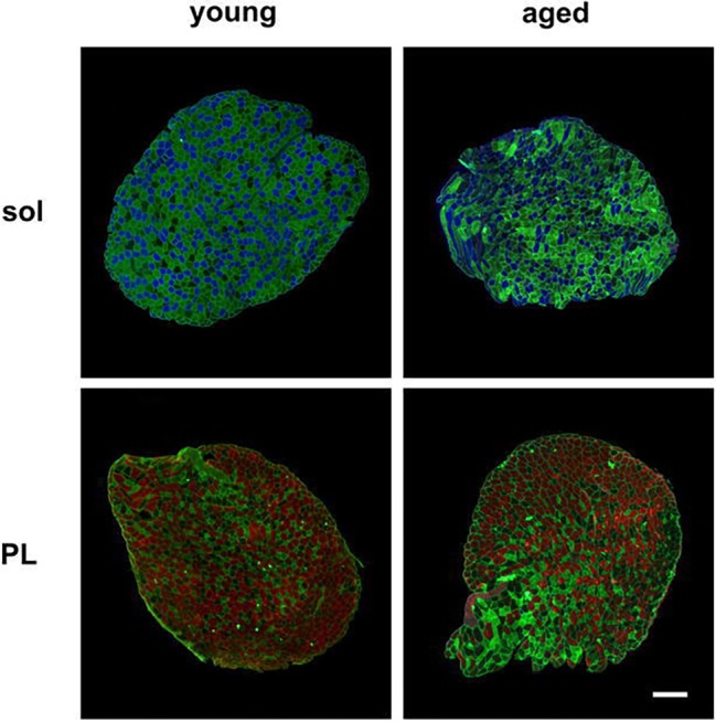
Figure 2.

Representative images of muscle-fiber typing of soleus and plantaris muscles. Muscle sections were stained with antibodies directed against myosin heavy chains. Indirect immunofluorescent staining of type I (blue), type IIb (red), type IIa (green), and type IId/x (no staining) fibers is shown in the soleus (sol) and plantaris (PL) muscles of the young and aged mice. Scale bar: 200 μm.
