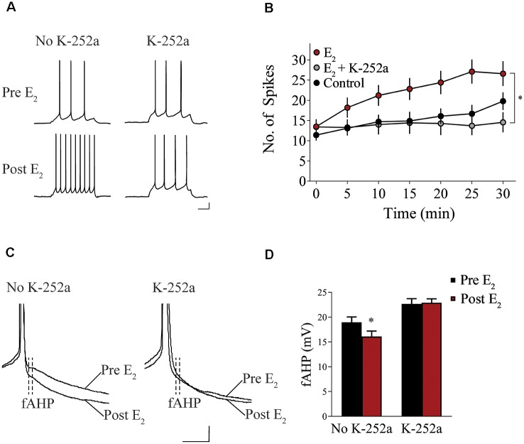
Figure 3.
Blockade of tropomyosin-related kinase (Trk) receptors prevents E2-induced potentiation of IL-mPFC neuronal intrinsic excitability. (A) Individual traces of current-evoked APs from IL-mPFC neurons that were not incubated in K-252a (left) or were incubated in K-252a (right), before (top) and after (bottom) bath-application of E2. Scale bars, 10 mV (vertical) and 500 ms (horizontal). (B) IL-mPFC neurons treated with E2 (n = 10) have increased intrinsic excitability compared to aCSF alone (n = 10) or neurons that were incubated in K-252a prior to bath-application of E2 (n = 7). (C) Representative waveforms showing fast afterhyperpolarization (fAHP) in neurons that were not incubated in K-252a (left) or were incubated in K-252a (right), and after bath-application of E2. Scale bars, 10 mV (vertical) and 10 ms (horizontal). (D) E2 suppresses fAHP and this effect was blocked in the presence of Trk receptor antagonist, K-252a. *p < 0.05. Error bars indicate SEM.

