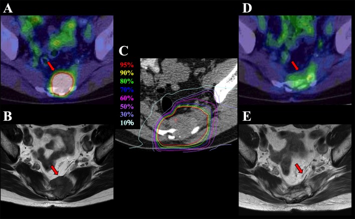
Figure 1.
Pelvic recurrence of rectal cancer in a 58-year-old man treated with C-ion RT. (A) PET before treatment. (B) MRI before treatment. (C) Dose distribution on axial CT images. Highlighted are: 95% (red), 90% (yellow), 80% (green), 70% (blue), 60% (pink), 50% (purple), 30% (light purple), and 10% (light blue) isodose curves [100% = 73.6 Gy [RBE]]. (D) PET 3 months after treatment. (E) MRI 12 months after treatment demonstrating disappearance of the presacral mass. Arrows show the recurrent tumor. C-ion RT, carbon-ion radiotherapy; PET, positron emission tomography; MRI, magnetic resonance imaging; CT, computed tomography; RBE, relative biological effectiveness.

