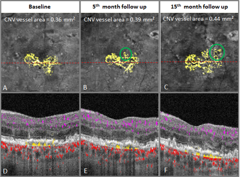
Figure 2.

Slow enlargement of non-exudative choroidal neovascularization (CNV) over 15 months. 3X3 mm en face outer retinal OCTA displayed over en face structural OCT (A-C) with CNV flow highlighted with yellow. Baseline (A) en face OCTA with subfoveal CNV. Emerging vascular branches (green circles) at 5 (B) and 15 (C) month follow-up visit. Cross-sectional OCTA (D-F) demonstrated flow between Bruch’s membrane and retinal pigment epithelium (yellow) without development of exudation.
