
Figure 3.
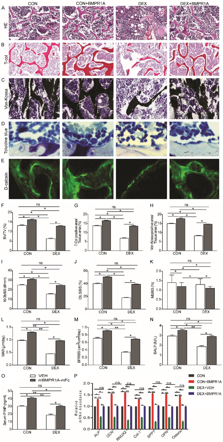
mBMPR1A-mFc treatment protects against the DEX-induced reduction of osteoblastic bone formation in mice. The histomorphometric analysis of distal femur by (A) hematoxylin & eosin (H&E), 400×, (B) total collagen, 400×, (C) von kossa, 400×, (D) toluidine blue, 400×, and (E) fluorescent calcein labeling staining, 400×. Histomorphometric analysis of (F) trabecular bone volume (BV/TV), (G) Total-Col positive area/Tissue area, (H) von kossa positive area/Tissue area, (I) Number of OBs per bone surface (N.Ob/BS), (J) Percentage of bone surfaces covered by OBs (Ob.S/BS), (K) percentage of bone surfaces covered by mineralized surfaces (MS/BS), (L) Mineral apposition rate (MAR), (M) Bone formation rate/bone surface (BFR/BS). Serum bone formation markers (N) BALP and (O) PINP were measured by ELISA. (P) The expression of osteoblast-specific genes, including ALP, OCN, Runx2, COL-1, SPP1, OPN and Osterix was examined by qRT-PCR in femurs. Values are expressed as mean ± SD, n=10 per group. ns, not statistically significant; *, P<0.05; **, P<0.01; ***, P<0.001, versus the indicated group.