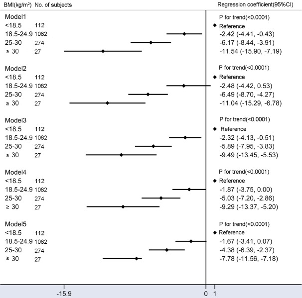
Figure 3.
Mean differences in fetal fraction according to maternal BMI (kg/m2). Model 1: Crude model. Model 2 were adjusted for GA and multiple gestations. Model 3 added average size of cell-free DNA on the basis of model 2. Model 4 added maternal age, maternal plasma cell-free DNA concentration, library concentration, and uniquely mapped reads on the basis of model 2. Model 5 added average size of cell-free DNA on the basis of model 4. Compared to models 2 and 4, models 3 and 5 included additional adjustments for confounding factors of the average size of cell-free DNA and significantly reduced fetal fraction differences between obese and underweight pregnant women.

