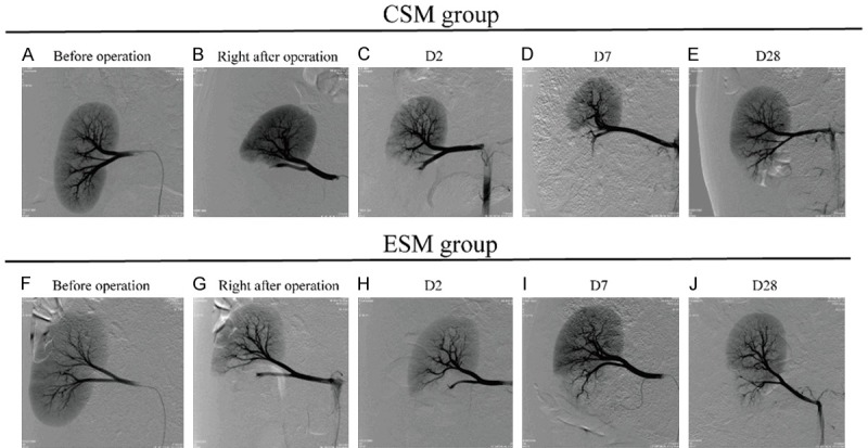
Figure 3.

The angiogram before the operation, right after the operation, on D2, D7 and D28 after the operation in CSM group and ESM group. Before operation, the staining of blood vessel and renal parenchyma could be clearly seen in CSM group (A) and ESM group (F). Right after the operation, the staining of the renal parenchyma and the blood vessel in lower pole were disappeared in CSM group (B) and ESM group (G). On D2 after treatment, the target arteries were embolized persistently in two groups (C, H). On D7 after treatment, the blood flow was partly reinstated in proximal arteries (D, I), and on D28 after treatment, the blood flow was partly reinstated in a proportion of distal arteries (E, J). CSM, CalliSpheres microspheres; ESM, Embosphere microspheres.
