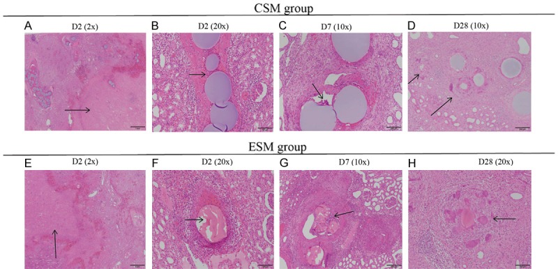
Figure 5.

Histopathology findings of the lower pole in CSM group and ESM group on D2, D7 and D28 after the operation. On D2 after the operation, there was cortical necrosis with hemorrhage/congestion in the periphery rim and congestion in medulla in CSM group (arrows) (A) and ESM group (E). Besides, inflammatory cells infiltrated in the perivascular area and there existed thrombosis in both groups (arrows) (B, F). The ESM was fragmented and incomplete in shape, which was most likely to be an artifact generated during slides preparation. On D7 after the operation, the microspheres were surrounded by multinucleated giant cells (arrows) in both groups (C, G). On D28 after the operation, microspheres were phagocytized by multinucleated giant cells in CSM group (D) and ESM group (arrows) (H). Specimen samples were examined using hematoxylin-eosin (H-E) staining. CSM, CalliSpheres microspheres; ESM, Embosphere microspheres; H-E, hematoxylin-eosin.
