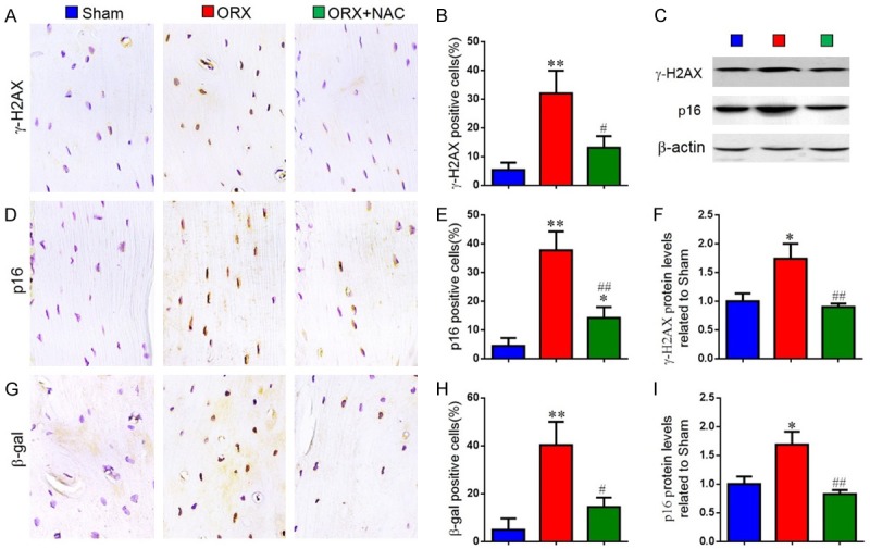
Figure 5.

The effect of NAC on DNA damage and osteocyte senescence in ORX mice. Representative micrographs of paraffin sections of tibias from 16-week-old mice of each group stained immunohistochemically for (A) γ-H2AX, (D) p16INK4a and (G) β-gal. The percentages of (B) γ-H2AX-positive, (E) p16INK4a-positive and (H) β-gal-positive cells were determined by image analysis. (C) Representative western blots of bone tissue extracts showing expression of γ-H2AX and p16INK4a; β-actin was used as loading control for the western blots in the sham, ORX and ORX+NAC groups. (F) γ-H2AX and (I) p16INK4a protein levels relative to β-actin protein levels were assessed by densitometric analysis and expressed relative to levels in sham-operated mice. Data are presented as the mean ± SEM of determinations, each data-point was the mean of five specimens. *P < 0.05, **P < 0.01 versus sham mice. #P < 0.05, ##P < 0.01 versus ORX mice. (A, D, G) Magnification, 400×.
