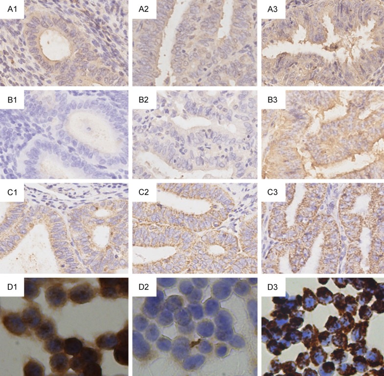
Figure 7.

Representative pictures of KLK6, KLK7, and KLK8 expression detected by IHC. A1-A3. Immunoperoxidase (DAB) labelling of KLK6 in EC tissue with Grades 1-3. B1-B3. Immunoperoxidase labelling of KLK7 in EC tissue with Grades 1-3. C1-C3. Immunoperoxidase labelling of KLK8 in EC tissue with Grades 1-3. D1-D3. Immunoperoxidase labelling of KLK6, 7, 8 in RL 95-2 cell line. Magnification was 200× for all pictures.
