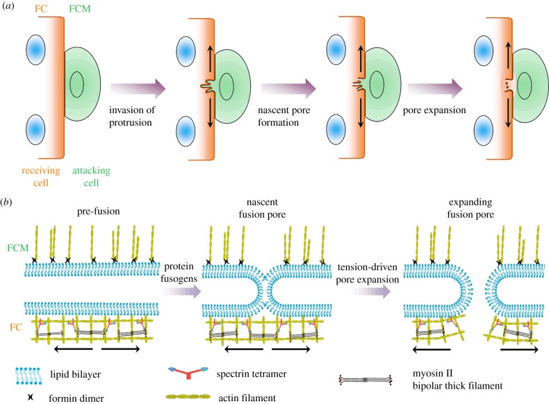
Figure 1.
Schematic diagram of the cell–cell fusion process. (a) The fusion process starts with the formation of multiple invasive protrusions in a fusion-competent cell, followed by the formation and expansion of fusion pores. (b) At the tip of the protrusion, the tension in the actin cytoskeleton facilitates the pore expansion during cell–cell fusion. The ‘attacking cell’ and ‘receiving cell’ refer to the FCM and FC, respectively. The arrows indicate the tension in the actin cytoskeleton underneath the plasma membrane. (Online version in colour.)

