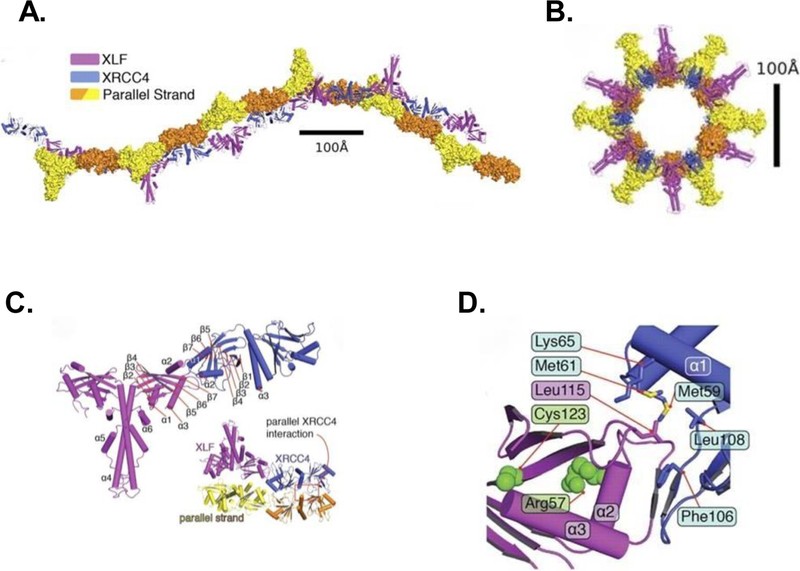
Figure 2.
Structure of the XLF-XRCC4 filament as determined by crystallography. A. Two orthogonal views of the biological unit (one helical turn) of the filament formed by XLF(1–224) (magenta) and XRCC4(1–140) (blue), as seen in the crystal lattice. Parallel strands (yellow/orange) of a second filament are in surface representation. B. View of the biological unit rotated by 90°. C. Overall architecture of the XLF(1–224)-XRCC4(1–140) complex, colored magenta and blue, respectively. Helices α4-α6 correspond to helices αD-αF in Figure 1. Inset shows interaction between XRCC4 dimers in parallel filaments. D. Interface between head domains of XLF(1–224) (magenta) and XRCC4(1–140) (blue). Leu115 of XLF occupies a hydrophobic pocket formed by XRCC4 residues Leu108, Phe106, Met59, Met61, and Lys65. (Image reproduced with permission from [24].)

