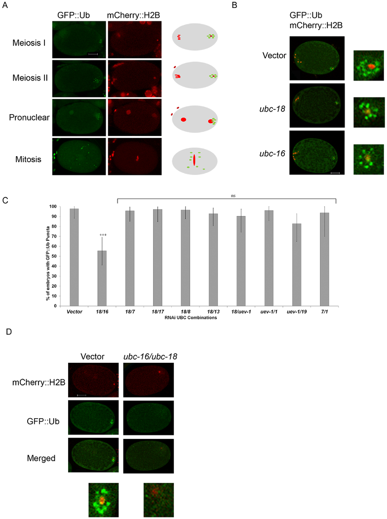
Figure 1. Simultaneous knockdown of ubc-18 and ubc-16 reduces ubiquitination of membraneous organelles.
Transgenic C. elegans embryos expressing GFP::Ub and mCherry::H2B were used to track the ubiquitination of MOs after RNAi knockdown of individual UBCs and double UBC combinations. (A) Wild type (vector control) embryos show the normal distribution of GFP::Ub following fertilization. The cartoon embryos to the right depict the characteristic pattern of GFP::Ub (green) and histone (red). In meiosis I and meiosis II embryos, GFP::Ub is concentrated on the MOs and localizes around the sperm DNA in the posterior region of the embryo (posterior is to the right). During pronuclear formation and pronuclear movement, the MOs remain ubiquitinated and become more dispersed in the embryo. During mitosis, the MOs are dispersed throughout with a tendency to localize near the midline of the embryo. (B) Embryos from worms fed RNAi bacteria of ubc-16 and ubc-18 singly did not show a reduction in GFP::Ub surrounding the paternal DNA. Embryos shown are meiosis I or meiosis II embryos. (C) RNAi screen of double knockdown of UBCs. The graph represents the top 9 hits from the UBC combination screen. All other RNAi combinations showed GFP::Ub around sperm DNA in more than 80% of embryos. Simultaneous knockdown of ubc-18 and ubc-16 was the most promising result from the screen. The double knockdown showed a 40% reduction in one cell embryos with GFP::Ub vesicles. A total of 20 embryos were observed for each condition and statistical differences were determined using a two tailed z test. ***p< 0.0001 (D) Representative embryos from the ubc-18/ubc-16 knockdown. Paternal DNA in embryos treated with ubc-18 and ubc-16 RNAi were not surrounded with GFP::Ub vesicles as is typically seen in empty vector treated worms. Scale bars 10 μm.

