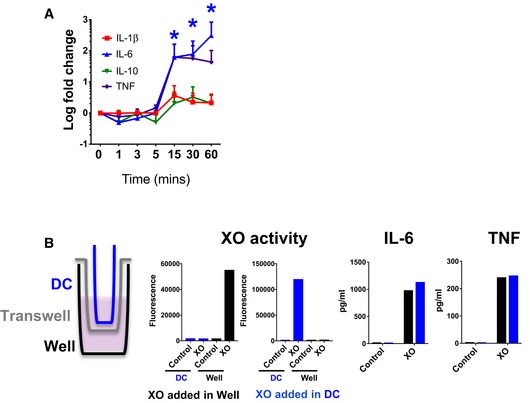
Figure 3. Prolonged exposure to extracellular ROS induces cytokine secretion.

-
AXO was incubated with macrophages for the indicated times, when wells were washed twice and media was replaced. Supernatants were collected for cytokine determinations 24 h later (n = 3). Error bars show standard deviation. Kruskal–Wallis statistical method was used for statistical analysis. Blue asterisks indicate significance when values were compared with time 0 (*P < 0.05).
-
BDiagram shows experimental set up with macrophages in the bottom of the well and XO added either in the dialysis cassette (DC, blue) or the well (black). XO activity panels show that the enzyme did not leak through the DC membrane. Secretion of IL‐6 and TNF was measured 24 h after addition of XO in each compartment. A representative result of two independent experiments is shown.
Source data are available online for this figure.
