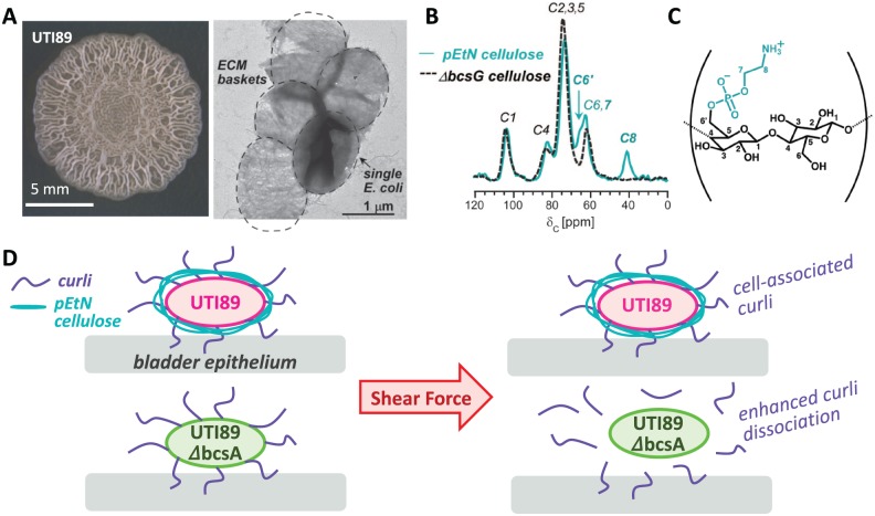
Figure 1.
(A) Two-day-old UTI89 Escherichia coli macrocolony biofilm on YESCA agar and transmission electron micrograph of extracted UTI89 ECM.6 (B) 13C CPMAS solid-state nuclear magnetic resonance spectra of pEtN cellulose and of the cellulosic component isolated from UTI89 ΔbcsG.9 The peaks at 41 and 63 ppm correspond to the C-8 and C-7 in the phosphoethanolamine functional group, respectively. The C-6 is shifted from 62 to 66 ppm in the pEtN cellulose. (C) Plausible representation of pEtN cellulose with alternating glucose and pEtN glucose, although modification patterning still must be determined. (D) Model depicting the role of pEtN cellulose in facilitating E coli attachment to bladder epithelium.12 pEtN cellulose acts as a type of glue to enhance curli association at the bacterial surface. ECM indicates extracellular matrix.

