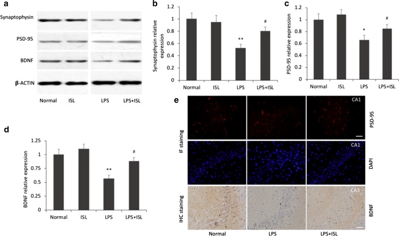
Fig. 2.
Effects of ISL on LPS-induced synaptic dysfunction. a–d Western blotting and histograms show the protein levels of synaptophysin, PSD-95 and BDNF in the hippocampus of each group. β-Actin was used as a loading control. Values are presented as mean ± SEM (n = 6). *p < 0.05 and **p < 0.01 vs. normal group, #p < 0.05 vs. LPS group. e Representative images show the immunoreactivities of PSD-95 in the CA1 subfield of the hippocampus by IF staining and BDNF in the CA3 subfield by IHC staining. Scale bars = 50 μm

