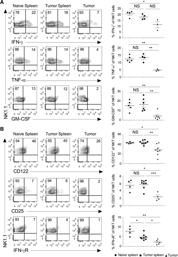
Fig. 2.
Intratumoral NKT cells show altered expression of cytokines and cytokine receptors. Naïve C57BL6 mice were given s.c. injection of B16F10 cells (1 X 10⁶ cells/mouse). a At day 13, spleen and tumors were harvested. The single cell suspension was stimulated with PMA/ionomycin, and intracellular cytokines expression was analyzed after gating on NKT cells. The representative contour plots are shown (left panel), and data from all the mice are shown (right panel). n = 3–6 mice/group. b On day 13, the surface expression of CD25 (IL-2Rα), CD122 (IL-2Rβ) and IFN-γR on NKT cells were analyzed (left). n = 4–8 mice/group. The bar represents s.e.m., and each dot represents data from an individual mouse (a, b). Student’s t-test (a, b). In all panels, *p < 0.05; **p < 0.01; *** p < 0.001; NS, not significant

