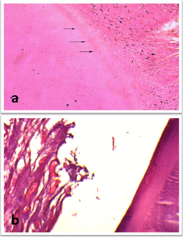
Fig. 4.

(a) Photomicrograph for a sample of subgroup B (MTA) at 2 month showing formation of new layer of dentin like tissue (arrows) on the inner root wall (H&E, X200). (b) Higher magnification of a sample of subgroup A (propolis) showing fibrous connective tissue with islands of cementum like tissue inside the root canal space (H&E, X200)
