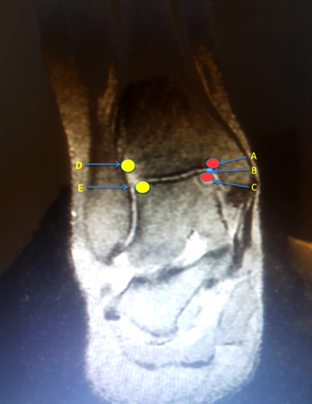
Figure 2.

The injection points. A) Medial joint surface of tibia. B) Intraarticular injection point. C) Medial joint surface of talus. D) Lateral joint surface of tibia. E) Lateral joint surface of talus. Injection point A, B, and C used for posteromedial lesions; Injection points B, D, and E used for anterolateral lesions.
