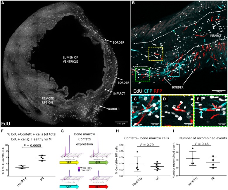
Figure 2.
A complete transverse section of the left ventricle at 7 days post-MI showing increased density of EdU expressing cells in the border region compared with the infarct and remote region (A). Dense neovascularization due to clonal proliferation of Pdgfb-lineage endothelial cells was observed in the infarct border region (B, with high power inserts in C–E). Confetti+Pdgfb-lineage endothelial cells frequently co-expressed EdU (B–E) and were significantly increased in the infarct border at 7 days post-MI compared with the Confetti+ EdU+ Pdgfb-lineage endothelial cells in the left ventricle of healthy uninjured mice (28.5 ± 4.8% vs. 58.5 ± 7.6%, P = 0.0005) (F). Representative flow cytometry plots showing very low reporter fluorophore expression in femoral bone marrow cells from adult Pdgfb-iCreERT2-R26R-Brainbow2.1 mice are shown in (G) with threshold gates set for each fluorophore using C57Bl6 wild type mice bone marrow cells as a negative control. No change in fluorophore expression by bone marrow cells was observed between healthy and MI groups (0.04 ± 0.02% vs. 0.03 ± 0.009%, P = 0.79) (H). The founding number of recombined events (where a Confetti+ cell or clone was counted as one event) was unchanged between healthy and MI groups (72.3 ± 9.0 vs. 67.3 ± 6.6 events per section, P = 0.46) (I).

