

Abstract
Aim:
This study aims to test the tensile strength and growth of Candida albicans on Viscogel tissue conditioner when incorporated with coconut oil (CCO) and to compare its efficacy with other antifungal agents.
Settings and Design:
Evaluative - In-vitro study design.
Materials and Methods:
Fifty dumbbell-shaped samples (n = 10) of Viscogel tissue conditioner were fabricated according to ASTM standard and were classified into 5 groups (10% CCO, 30% w/w tea tree oil, 5% w/w fluconazole, 0.03% w/w silver nanoparticles, and plain tissue conditioner). These samples were compared and evaluated for their tensile strength. Further to test the antifungal activity, a total of 60 samples (n = 15) were fabricated, each group (n = 15) was further divided into three subgroups (n = 5), namely 24-h, 3-day, and 5-day period, which were inoculated in sabouraud dextrose agar plate to test for the growth of C. albicans.
Statistical Analysis Used:
Oneway ANOVA and post hoc Tukey honestly significant difference test.
Results:
10% w/w CCO yielded a mean tensile strength of 20.06 as compared to the plain tissue conditioner which showed a mean tensile strength of 17.81. Similarly, 10% w/w CCO incorporated into Viscogel tissue conditioner showed a significant reduction in the colonization of C. albicans on the 5th day.
Conclusions:
10% w/w of CCO when mixed with Viscogel tissue conditioner showed a significant reduction in the growth of C. albicans, and addition of the same increased the tensile strength of the tissue conditioner.
Keywords: Antifungal agents, Candida albicans, coconut oil, tensile strength, tissue conditioner
INTRODUCTION
As the access to dental care is improving, people are retaining their natural teeth for a longer period of time, but the incidence of edentulousness still remains significant. Removable dental prosthesis has been and remains the basic treatment for patients with complete or partial edentulousness. The need for long-term use of removable prosthesis can bring about profound changes in the oral cavity, and one such commonly encountered sequel of wearing removable prosthesis is denture stomatitis.[1]
Successful treatment plan for denture stomatitis includes elimination of the local irritating factors, treatment of the abused tissue, diet counseling, systemic evaluation, replacement of the old prosthesis, establishment of proper occlusal scheme and correction of any occlusal prematurities, meticulous maintenance of oral hygiene, and use of soft tissue liners.
Lately, tissue conditioners have been used as a drug delivery tool for elderly patients suffering from denture stomatitis caused due to Candida infection, as the success of topical application of drugs in the oral cavity may be compromised by lack of patient compliance.[2,3] Furthermore, maintaining the regular and optimal dose of topical antifungal agents is challenging in geriatric denture wearers due to reduced motor skills, cognitive impairment, and memory loss.[4] To overcome these difficulties, antifungal agents have been incorporated into soft tissue liners and are checked for their effectiveness against Candida albicans.
There are many published reports on antifungal agents such as nystatin, azole group derivatives, chlorhexidine, metallic oxide powders, photocatalysts, and silver nanoparticles incorporated into tissue conditioners, which have been reported to have varying degree of success.[1,5,6,7,8,9,10,11,12,13,14,15] However, due to the resistance developed by Candida species toward these drugs, therapeutic effects of plant oils that have antifungal properties are being explored.
A number of studies have been carried out which have investigated the effect of natural and herbal antimicrobials against C. albicans by incorporating them into tissue conditioners, for example, tea tree oil (TTO), lemongrass oil, thai herbs, and origanum oil.[16,17,18,19,20,21]
One such herbal oil that has both antifungal and antimicrobial property is Cocos nucifera or commonly known as coconut. Coconut oil (CCO) has been proven to have antimicrobial and antifungal property against Candida species including C. albicans, Candida glabrata, Candida tropicalis, Candida parapsilosis, and Candida krusei.[22]
What makes CCO unique as compared to other oils is the presence of medium chain fatty acids as compared to the long chain fatty acids present in other edible oils. CCO has 92% saturated fatty acids, of which 50% is lauric acid. It has been postulated that lauric acid along with other medium chain fatty acids has the capacity to modify the cell wall of microorganisms, disrupt their cellular mechanism by infiltrating the cell membrane, and obstruct the enzymes involved in energy production and nutrient transfer, leading to the death of the microorganism.[23]
Thus, the purpose of this in vitro study was to analyze the effect of CCO on C. albicans by incorporating it into tissue conditioners and compare its efficacy with TTO, silver nano particles (SNPs), and fluconazole (FLU).
The null hypothesis was that there will be no antifungal activity exhibited toward C. albicans and no change in the tensile strength of Viscogel tissue conditioner when incorporated with CCO.
MATERIALS AND METHODS
Viscogel tissue conditioner incorporated with CCO, TTO, SNPs, and FLU were used as samples [Table 1].
Table 1.
Materials used in the study
| Type | Material | Concentration (%) | Code | Manufacturer |
|---|---|---|---|---|
| Tissue conditioner | Viscogel tissue conditioner | - | - | DENTSPLY DeTrey GmbH, Germany |
| Antifungal agent | Coconut oil | 10 | CCO | Falcon, Bangalore, India |
| Antifungal agent | Tea tree oil | 30 | TTO | Falcon, Bangalore, India |
| Antifungal agent | Fluconazole | 5 | FLU | Aurobindo Pharma Ltd, Hyderabad, India |
| Antifungal agent | Silver nanoparticles | 0.03 | SNP | Aadarsh innovations, Pune, India |
Methodology to determine the optimum concentration of coconut oil
A pilot study was performed to determine the optimum concentration of CCO, at which the antifungal testing could be carried out without altering the physical properties of the tissue conditioner. The pilot study involved incorporating four different concentrations of CCO into the tissue conditioner and evaluating its effect on its physical property. The concentration which yielded the maximum tensile strength under maximum load was preferred for the antifungal study.
Preparation of mold
A total of 12 wax samples (n = 3) were fabricated using modeling wax (Hindustan Modeling Wax No. 2, The Hindustan Dental Products, Hyderabad, India) according to the ASTM standard (ASTM D412) with a cross-sectional area of 20 mm × 6 mm × 3 mm. These wax samples were invested in the lower part of the flask with type IV dental stone or die stone (Kalrock; Kalabhai Karson Pvt, Ltd), flasked, and dewaxed to form molds.
Preparation of samples to test tensile strength
One control group of tissue conditioner with no CCO incorporated and three test groups with varying concentrations of CCO (10% w/w, 20% w/w, and 30% w/w) were added to 3 g of tissue conditioner powder and 2.2 ml of liquid, hand mixed in a plastic cup with a mixing spatula as per manufacturer's recommendation. The tissue conditioners were homogeneously manipulated and loaded into the lower part of the mold, flasked, and allowed to gel. The set samples were retrieved, and excess was trimmed with a Bard-Parker Knife [Figure 1].
Figure 1.
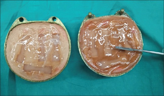
Preparation of samples to test tensile strength
The 12 samples (n = 3) thus obtained were categorized into 4 groups and stored in distilled water in 4 separate plastic beakers at 37°C for 7 days, which were changed regularly.
The samples were grouped as follows:
Group 1: Control group
Group 2: 10% CCO
Group 3: 20% CCO
Group 4: 30% CCO.
On the 7th day, the maximum tensile strength evaluation was carried out for all the groups using an Instron Universal testing machine (Model 3366, Instron Corp., Canton, Mass.) at a rate of 40 mm/min. The tissue conditioner with 10% w/w of CCO exhibited a mean tensile strength of 23.31 as compared with the 20% w/w and 30% w/w concentration of CCO which showed a mean tensile strength of 18.78 and 14.00, respectively. Hence, 10% w/w of CCO was preferred for further antifungal testing.
Similarly, tensile strength evaluation and comparison of 10% w/w CCO with 5% w/w FLU, 30% w/w TTO, and 0.03% w/w SNP were carried out using the same method. A total of 50 samples (n = 10) were made, which were grouped as follows:
Group A: 10% w/w CCO
Group B: 30% w/w TTO
Group C: 5% w/w FLU
Group D: 0.03% w/w SNP
Group E: plain tissue conditioner which acted as a control group.
Preparation of samples to test antifungal effect
Preparation of mold
Thirty circular wax patterns 8 mm in diameter and 1.2 mm in cross-section were fabricated using modeling wax (Hindustan Modeling Wax No. 2, The Hindustan Dental Products, Hyderabad, India) and arranged on a glass slab. An impression of the arranged wax patterns was made using putty impression material (GC Flexceed). The obtained putty mold was utilized for making a stone model. A vacuum forming thermoplastic sheet (TJ dental, Mumbai, India) was placed over the stone mold and molded under vacuum, using a vacuum-forming unit (Dunaform, Al Dente). The formed thermoplastic sheet was retrieved and subsequently used as a mold to prepare the samples for microbiological testing.
Preparation of the samples
Mixing coconut oil with Viscogel tissue conditioner
10% w/w of CCO was hand mixed with Viscogel tissue conditioner according to manufacturer's instructions in a plastic mixing cup. With the help of a mixing spatula, the tissue conditioner mixture was applied over the thermoplastic sheet mold and allowed to gel. A total of 15 samples were made [Figure 2].
Figure 2.
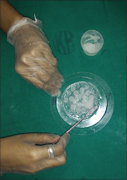
Preparation of samples for microbiological study
The set samples were placed into 2 ml cryovials which were labeled as follows:
1CCO1–1CCO5 = 5 samples for the 1st day culture
3CCO1–3CCO5 = 5 samples for the 3rd day culture
5CCO1–5CCO5 = 5 samples for the 5th day culture.
Similarly, 30% w/w of TTO, 5% w/w of FLU, and 0.03% SNP were mixed homogeneously with Viscogel tissue conditioner according to manufacturer's instructions and allowed to gel. The obtained samples were placed separately in 2 ml cryovials and labeled accordingly.
Therefore, a total of 60 samples (n = 15) were made, which were stored in 2 ml cryovials and kept for ultraviolet sterilization.
Preparation of inoculum
Standard strain of ATCC 24433 C. albicans was inoculated into Sabouraud Dextrose broth (Himedia Laboratories) and incubated at 37°C. After 8 h, C. albicans suspension was standardized by dilution with sterile broth to a density visually equivalent to barium sulfate standard 0.5 McFarland.
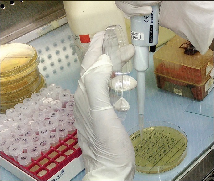
Inoculation of samples with Candida albicans
In 2 ml cryovials, 1 ml of Sabouraud dextrose agar broth was filled using the P1000 micropipette, and 10 μl of diluted C. albicans was dropped into each vial using a P10 micropipette. These vials were then stored in an incubator under 37°C for 1, 3, and 5 days.
Inoculation of agar plate using spot inoculation method
24-h inoculation
The agar plate was divided into a total of 20 boxes, i.e., 5 columns – one for each sample and 4 rows – one for each group of antifungal agent. 10 μl of C. albicans Sabouraud Dextrose Agar broth solution was pipetted out from the cryovials labeled 1CCO1–1CCO5, 1FLU1–1FLU5, 1SNP1–1SNP5, and 1TTO1–1TTO5 using a P10 micropipette and spot inoculated into each box [Figure 3]. The agar plate was sealed using Parafilm M sealing tape to prevent cross contamination and stored in an incubator at 37°C for 24 h. The plate was examined for growth of C. albicans after 24 h.
Figure 3.
Inoculation of agar plate using spot inoculation method
Similar procedure was carried out for the 3rd and 5th day inoculation by obtaining the inoculum from the labeled 3rd-day and 5th-day cryovials, respectively. Both the agar plates were examined after 24 h interval to evaluate the growth of C. albicans.
The 24-h, 3rd-day, and 5th-day agar plates were further compared for the effective antifungal property of each agent against C. albicans.
RESULTS
Study demonstrated 10% w/w of CCO to have the mean tensile strength of 23.31 as compared with the 20% w/w and 30% w/w concentration of CCO which showed a mean tensile strength of 18.78 and 14.00, respectively. Hence, 10% w/w of CCO was preferred for further antifungal testing.
Descriptive statistical analysis of mean tensile strength and standard deviation of each group in Table 2 demonstrates SNP to have a mean tensile strength of 21.63, followed by CCO which shows a mean tensile strength of 20.06, FLU showing 18.55 and plain tissue conditioner exhibiting a mean tensile strength of 17.81. TTO exhibited the least mean tensile strength of 10.58.
Table 2.
Descriptive statistics of tensile strength and standard deviation of each group
| Group | n | Mean | SD | SE | Minimum | Maximum |
|---|---|---|---|---|---|---|
| A | 10 | 20.0600 | 2.03740 | 0.64428 | 17.44 | 23.01 |
| B | 10 | 10.5800 | 0.80954 | 0.25600 | 9.52 | 11.72 |
| C | 10 | 18.5590 | 2.47627 | 0.78306 | 16.00 | 22.55 |
| D | 10 | 21.6350 | 2.60470 | 0.82368 | 18.62 | 25.50 |
| E | 10 | 17.8170 | 0.94701 | 0.29947 | 16.85 | 19.35 |
| Total | 50 | 17.7302 | 4.26789 | 0.60357 | 9.52 | 25.50 |
SD: Standard deviation, SE: Standard error
Hence, adding 10% w/w of CCO to Viscogel tissue conditioner increases its tensile strength as compared to that of plain Viscogel tissue conditioner.
One-way ANOVA analysis followed by post hoc Tukey honestly significant difference test was carried out between the groups for assessing the significant difference between any of the five groups [Tables 3 and 4]. The significance level was set at P < 0.05. There was no statistical difference between the test group, i.e., 10% CCO with 5% w/w FLU and 0.03% w/w SNP (P > 0.05), whereas there was a significant difference between the 10% CCO and 30% w/w TTO (P < 0.05).
Table 3.
One-way ANOVA analysis
| Sum of squares | df | Mean square | F | Significance | |
|---|---|---|---|---|---|
| Between groups | 724.952 | 4 | 181.238 | 48.669 | 0.000 |
Table 4.
Post hoc Tukey honestly significant difference analysis to statistically compare the tensile strength between Groups A, B, C, D, and E
| Group (I) | Group (J) | Mean difference (I-J) | SE | P |
|---|---|---|---|---|
| A | B | 9.48000* | 0.86301 | 0.000 |
| C | 1.50100 | 0.86301 | 0.421 | |
| D | −1.57500 | 0.86301 | 0.372 | |
| E | 2.24300 | 0.86301 | 0.088 |
*P<0.05 significance difference. SE: Standard error
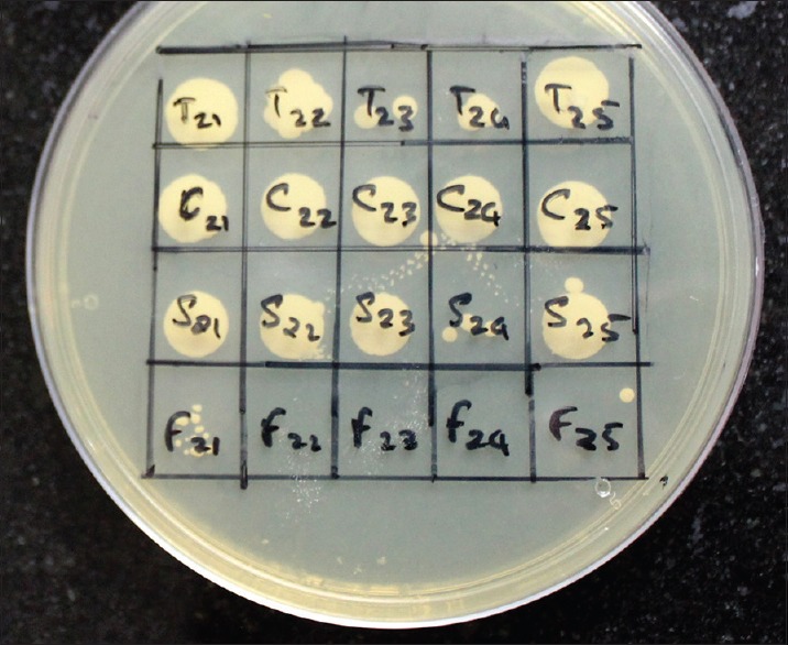
The microbiological study carried out to determine the antifungal activity of Viscogel tissue conditioner against C. albicans when incorporated with 10% w/w of CCO, displayed a gradual and steady decrease in the growth of C. albicans when incubated for 24 h, 3 days, and 5 days [Figures 4-6].
Figure 4.
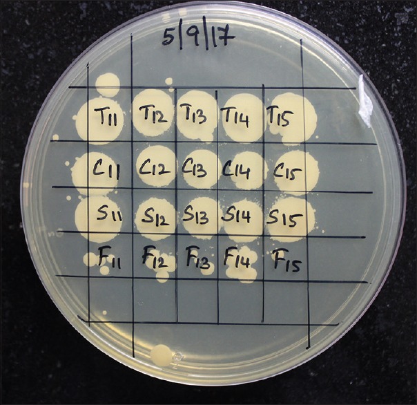
Evaluation of growth of Candida albicans after 24 h of inoculation
Figure 6.
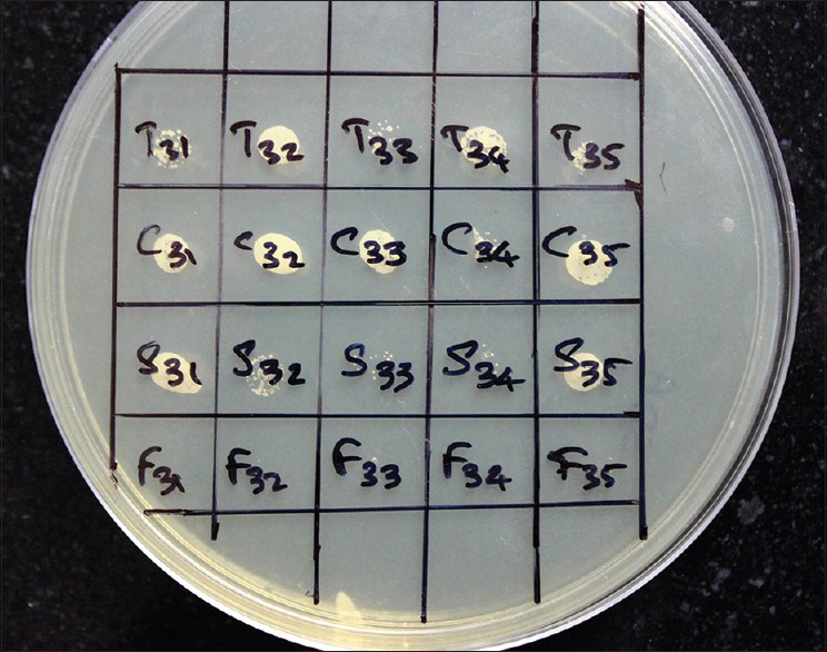
Evaluation of growth of Candida albicans on 7th day of inoculation
Figure 5.
Evaluation of growth of Candida albicans on 3rd day of inoculation
DISCUSSION
Denture stomatitis is an opportunistic infection related to an inflammatory process which compromises the mucosal surface beneath the dentures. Although the etiology is multifactorial, C. albicans is the only microorganism with an established pathogenic role.
A possible initial step in the treatment of denture stomatitis is advising the patient to wear dentures only for few hours in a day; however, this is not practical for the patient. Due to its multifactorial etiology, the management of Candida-associated denture stomatitis is also intricate. Topical application of antifungal drugs can be ineffective as it depends on patient compliance. Hence, tissue conditioners incorporated with antifungal agents have been into use as an antifungal drug delivery system to treat the abused tissue under the denture-bearing area. This can be a promising method of drug delivery system to overcome the obstacles of other therapies.
The most commonly used combination of antifungal agent and tissue conditioner in literature is nystatin and Viscogel. Nystatin when incorporated into tissue conditioners has shown antifungal and antimicrobial activity, but its effect was comparatively less as compared with the azole groups.[1,7,10,12]
Another commonly used antifungal agent is Amphotericin B. Although it is a good and potent antifungal agent, when incorporated into tissue conditioners, it is completely ineffective.[14,24] This could be due to the inability of the compound to be stable or to convert its chemistry when incorporated into tissue conditioners.
Among the organic antimicrobial agents, azole group derivatives were found to be the most effective and potent against C. albicans.[1,7,10,12] Although they are found to be effective, prolonged usage may cause drug resistance, hepatotoxicity, and renal toxicity.[5,6,7,8,9,10] Furthermore, topical application of the same can result in unpleasant taste and poor patient compliance due to loss of memory or manual dexterity of the aged patient.
To avoid the complications of organic antimicrobial agents, researchers started incorporating inorganic compounds such as SNPs, magnesium oxide, photocatalysts, and silver zeolite into tissue conditioners, to test its antifungal activity against C. albicans.[15,16,17,18] It has been proven that silver zeolite when incorporated into soft liners has substantial effect against plaque-forming bacteria and also is not affected by human saliva.[18]
The advancement in nanotechnology has brought into use nanoparticles such as silver and gold. Due to their increased surface area and enhanced reactivity, they have been considered as potent antimicrobial agents.[19] SNPs have been incorporated into tissue conditioners, and it has been proven to prevent colonization of C. albicans.[19] However, the disadvantages of using SNPs include their high cost, discoloration of tissue conditioner when incorporated with the same, and metallic taste.
Photocatalyst like photocatalytic titania has been proven to have an antifungal effect when used as an antifungal coating on tissue conditioner.[20] However, according to Uchimaru et al., there has been a reduction in the antifungal activity after incorporating photocatalyst (Photohap) in tissue conditioner.[15]
In addition to these, a number of studies have been carried out which have investigated the effect of natural and herbal antimicrobial agents against C. albicans by incorporating them into tissue conditioners, which have shown good antifungal and antimicrobial activity.[22,23,25,26] The main advantage of using natural and herbal medicaments over organic and inorganic compounds is its ready availability, minimal or no side effects, and cost-effectiveness.
One such herbal oil that has antifungal and antimicrobial property but has not been incorporated into tissue conditioner to check its effectiveness against Candida-associated denture stomatitis is C. nucifera or commonly known as coconut.
It has been proven that 5% w/w CCO has substantial antifungal activity against C. albicans biofilm isolated from clinical samples, which showed a marked reduction under scanning electron microscopic examination.[27] Furthermore, in another study carried out by PS Kumar, it has been substantiated that incorporation of 20% v/v of CCO reduced the growth of C. albicans.[28] However, as incorporation of any substance into the polymeric structure of tissue conditioner can affect its physical property, in this study, various concentrations of virgin CCO (10% w/w, 20% w/w, and 30% w/w) were incorporated into Viscogel tissue conditioner and tested for its effect on the tensile strength of the tissue conditioner.[29]
The tensile strength of a relining material provides us with the information on the ultimate tensile strength of the material in tension.[30] Subjecting a rubber material to compression and shear stress does not provide us a guide to determine quality of the material; whereas, the tensile strength of a rubber material is a measure to determine its quality to resist rupture under wet environmental conditions. Soft liners especially loose their adhesion to denture base surface, loose resiliency, and also have accumulation of plaque and debris.
The results exhibited 10% w/w CCO to have the maximum mean tensile strength as compared with the 20% w/w and 30% w/w concentrations of CCO.
The descriptive statistical analysis between the groups demonstrated SNP to have the maximum mean tensile strength of 21.63 followed by CCO which showed a mean tensile strength of 20.06. It was also proved statistically that incorporating TTO into Viscogel tissue conditioner reduced its mean tensile strength drastically to 10.58 from 17.81.
All the samples were stored in distilled water simulating the wet environment provided by saliva. The wet environment allows the ethanol and esters to get leached into water, which is then absorbed by the polymeric phase of the gel. This may cause changes in the viscoelastic properties of tissue conditioner.[30]
For evaluation of antifungal activity against C. albicans, spot inoculation technique has been employed. Although disc diffusion and agar punch well methods have been used in previous studies, it can cause dehydration of the agar medium, as they have to be kept incubated at 37°C for 7 days. This could give a false-positive result of inhibition of growth of C. albicans on the agar medium. Furthermore, C. albicans requires continuous supply of nutrition for its growth. This condition was fulfilled by the spot inoculation technique used in this in vitro study, as the samples were inoculated in SDA (Sabouraud Dextrose Agar) broth along with diluted C. albicans, where the microorganism received continuous nutrition supply for its growth. Therefore, the chances of false-positive inhibition of C. albicans growth were minimal or negligible.
The results of the present microbiological study revealed that there was a gradual reduction in the colonization of C. albicans in all the antifungal agents, when compared between the 24-h incubation period and the 5th-day incubation period. Furthermore, FLU displayed a complete inhibition of growth of C. albicans on day 5. This clearly proves that leaching of tissue conditioner when incorporated with antifungal agents is slow, consistent, and gradual which was in accordance with Graham et al. who have stated in their in vitro study that tissue conditioners flow for a period of 7 days and that they are clinically effective throughout this period.[29]
Furthermore, results of tensile strength evaluation proved that Viscogel tissue conditioner when incorporated with CCO, SNP, and FLU showed significant increase in its tensile strength as compared with the plain Viscogel tissue conditioner; on the other hand, TTO when incorporated into Viscogel tissue conditioner demonstrated a drastic decrease in its tensile strength.
Since only one tissue conditioner was used in the study, the results obtained here may therefore not apply to other tissue conditioners incorporated with CCO. Studies by incorporating the same into different brands of tissue conditioner should be carried out, and further in vivo studies are required using the combination of CCO and tissue conditioners to treat denture stomatitis. Furthermore, additional studies are required to determine the rate of release of these antifungal agents from tissue conditioner.
CONCLUSIONS
10% CCO was effective in producing significant antifungal activity on C. albicans, and the antifungal activity improved gradually from 24 h to day 5. At the same time, incorporation of the same statistically increased the tensile strength of Viscogel tissue conditioner when tested, after 7 days. Thus, we suggest CCO when mixed with Viscogel tissue conditioner can be used against C. albicans resistant to conventional therapies and also that this association showed an increase in tensile strength of Viscogel.
Financial support and sponsorship
Nil.
Conflicts of interest
There are no conflicts of interest.
Acknowledgment
We would like to extend our sincere and heartfelt gratitude to Mr. Stalin Lobo, Post graduate, Department of Microbiology, KMC Manipal for taking out his time even during his examinations and helping us with the microbiological procedures for the study without which, this study would have been impossible.
REFERENCES
- 1.Chow CK, Matear DW, Lawrence HP. Efficacy of antifungal agents in tissue conditioners in treating candidiasis. Gerodontology. 1999;16:110–8. doi: 10.1111/j.1741-2358.1999.00110.x. [DOI] [PubMed] [Google Scholar]
- 2.Hashem MI. Advances in soft denture liners: An update. J Contemp Dent Pract. 2015;16:314–8. doi: 10.5005/jp-journals-10024-1682. [DOI] [PubMed] [Google Scholar]
- 3.Iqbal Z, Zafar MS. Role of antifungal medicaments added to tissue conditioners: A systematic review. J Prosthodont Res. 2016;60:231–9. doi: 10.1016/j.jpor.2016.03.006. [DOI] [PubMed] [Google Scholar]
- 4.Emami E, Kabawat M, Rompre PH, Feine JS. Linking evidence to treatment for denture stomatitis: A meta-analysis of randomized controlled trials. J Dent. 2014;42:99–106. doi: 10.1016/j.jdent.2013.11.021. [DOI] [PubMed] [Google Scholar]
- 5.Geerts GA, Stuhlinger ME, Basson NJ. Effect of an antifungal denture liner on the saliva yeast count in patients with denture stomatitis: A pilot study. J Oral Rehabil. 2008;35:664–9. doi: 10.1111/j.1365-2842.2007.01805.x. [DOI] [PubMed] [Google Scholar]
- 6.Falah-Tafti A, Jafari AA, Lotfi-Kamran MH, Fallahzadeh H, Hayan RS. A comparison of the efficacy of nystatin and fluconazole incorporated into tissue conditioner on the in vitro attachment and colonization of Candida albicans. Dent Res J (Isfahan) 2010;7:18–22. [PMC free article] [PubMed] [Google Scholar]
- 7.Julian J, Sunil S, Baby GG. Efficacy of tissue conditioner acting as effective fungicidal drug delivery system – An in vitro study. Oral Maxillofac Pathol J. 2010;1:10–5. [Google Scholar]
- 8.Chopde N, Pharande A, Khade MN, Khadtare YR, Shah SS, Apratim A. In vitro antifungal activity of two tissue conditioners combined with nystatin, miconazole and fluconazole against Candida albicans. J Contemp Dent Pract. 2012;13:695–8. doi: 10.5005/jp-journals-10024-1211. [DOI] [PubMed] [Google Scholar]
- 9.Fallah-Tafti A, Jafari A, Mirzaeiipoorm L, Ashoori H. Stability and duration of antifungal effects of nystatin and fluconazole mixed with a tissue conditioner on colonization of Candida albicans (in vitro) J Res Dent Sci. 2014;11:21–6. [Google Scholar]
- 10.Bueno MG, Urban VM, Barbério GS, da Silva WJ, Porto VC, Pinto L, et al. Effect of antimicrobial agents incorporated into resilient denture relines on the Candida albicans biofilm. Oral Dis. 2015;21:57–65. doi: 10.1111/odi.12207. [DOI] [PubMed] [Google Scholar]
- 11.Gupta H, Bhat A, Prasad K, Kumar V. An innovative method of incorporating antifungal agents into tissue conditioners: An in vitro study. Trends Biomater Artif Organs. 2011;25:63–6. [Google Scholar]
- 12.Radnai M, Whiley R, Friel T, Wright PS. Effect of antifungal gels incorporated into a tissue conditioning material on the growth of Candida albicans. Gerodontology. 2010;27:292–6. doi: 10.1111/j.1741-2358.2009.00337.x. [DOI] [PubMed] [Google Scholar]
- 13.Schneid TR. An in vitro analysis of a sustained release system for the treatment of denture stomatitis. Spec Care Dentist. 1992;12:245–50. doi: 10.1111/j.1754-4505.1992.tb00458.x. [DOI] [PubMed] [Google Scholar]
- 14.Kanathila H, Bhat AM, Krishna PD. The effectiveness of magnesium oxide combined with tissue conditioners in inhibiting the growth of Candida albicans: An in vitro study. Indian J Dent Res. 2011;22:613. doi: 10.4103/0970-9290.90324. [DOI] [PubMed] [Google Scholar]
- 15.Uchimaru M, Sakai T, Moroi R, Shiota S, Shibata Y, Deguchi M, et al. Antimicrobial and antifungal effects of tissue conditioners containing a photocatalyst. Dent Mater J. 2011;30:691–9. doi: 10.4012/dmj.2010-203. [DOI] [PubMed] [Google Scholar]
- 16.Nam KY. In vitro antimicrobial effect of the tissue conditioner containing silver nanoparticles. J Adv Prosthodont. 2011;3:20–4. doi: 10.4047/jap.2011.3.1.20. [DOI] [PMC free article] [PubMed] [Google Scholar]
- 17.Matsuura T, Abe Y, Sato Y, Okamoto K, Ueshige M, Akagawa Y. Prolonged antimicrobial effect of tissue conditioners containing silver-zeolite. J Dent. 1997;25:373–7. doi: 10.1016/s0300-5712(96)00050-4. [DOI] [PubMed] [Google Scholar]
- 18.Morones JR, Elechiguerra JL, Camacho A, Holt K, Kouri JB, Ramírez JT, et al. The bactericidal effect of silver nanoparticles. Nanotechnology. 2005;16:2346–53. doi: 10.1088/0957-4484/16/10/059. [DOI] [PubMed] [Google Scholar]
- 19.Chladek G, Mertas A, Barszczewska-Rybarek I, Nalewajek T, Zmudzki J, Król W, et al. Antifungal activity of denture soft lining material modified by silver nanoparticles-a pilot study. Int J Mol Sci. 2011;12:4735–44. doi: 10.3390/ijms12074735. [DOI] [PMC free article] [PubMed] [Google Scholar]
- 20.Akiba N, Hayakawa I, Keh ES, Watanabe A. Antifungal effects of a tissue conditioner coating agent with TiO2 photocatalyst. J Med Dent Sci. 2005;52:223–7. [PubMed] [Google Scholar]
- 21.Budtz-Jörgensen E. The significance of Candida albicans in denture stomatitis. Scand J Dent Res. 1974;82:151–90. doi: 10.1111/j.1600-0722.1974.tb00378.x. [DOI] [PubMed] [Google Scholar]
- 22.Sharma S, Hegde V. Comparative evaluation of antifungal activity of melaleuca oil and fluconazole when incorporated in tissue conditioner: An in vitro study. J Prosthodont. 2014;23:367–73. doi: 10.1111/jopr.12117. [DOI] [PubMed] [Google Scholar]
- 23.Choonharuangdej S, Amornvit P, Srithavaj T, Alam MK. In vitro anti-Candida effect of Thai herbs supplemented in tissue conditioner. Int Med J. 2014;21:331–34. [Google Scholar]
- 24.Thomas CJ, Nutt GM. The in vitro fungicidal properties of visco-gel, alone and combined with nystatin and amphotericin B. J Oral Rehabil. 1978;5:167–72. doi: 10.1111/j.1365-2842.1978.tb01210.x. [DOI] [PubMed] [Google Scholar]
- 25.Srivatstava A, Ginjupalli K, Perampalli NU, Bhat N, Ballal M. Evaluation of the properties of a tissue conditioner containing origanum oil as an antifungal additive. J Prosthet Dent. 2013;110:313–9. doi: 10.1016/S0022-3913(13)60381-9. [DOI] [PubMed] [Google Scholar]
- 26.Catalán A, Pacheco JG, Martínez A, Mondaca MA. In vitro and in vivo activity of Melaleuca alternifolia mixed with tissue conditioner on Candida albicans. Oral Surg Oral Med Oral Pathol Oral Radiol Endod. 2008;105:327–32. doi: 10.1016/j.tripleo.2007.08.025. [DOI] [PubMed] [Google Scholar]
- 27.Agarwal V, Lal P, Pruthi V. Prevention of Candida albicans biofilm by plant oils. Mycopathologia. 2008;165:13–9. doi: 10.1007/s11046-007-9077-9. [DOI] [PubMed] [Google Scholar]
- 28.Kumar PS. Comparative evaluation of efficacy of antifungal activity of Azadirachta indica, Melaleuca oil and Cocos nucifera oil against Candida albicans incorporated in soft relining materials. J Indian Prosthodont Soc. 2018;18(Suppl S2):69. [Google Scholar]
- 29.Graham BS, Jones DW, Sutow EJ. Clinical implications of resilient denture lining material research. Part II: Gelation and flow properties of tissue conditioners. J Prosthet Dent. 1991;65:413–8. doi: 10.1016/0022-3913(91)90234-n. [DOI] [PubMed] [Google Scholar]
- 30.Garg A, Shenoy KK. A comparative evaluation of effect on water sorption and solubility of a temporary soft denture liner material when stored either in distilled water, 5.25% sodium hypochlorite or artificial saliva: An in vitro study. J Indian Prosthodont Soc. 2016;16:53–62. doi: 10.4103/0972-4052.167931. [DOI] [PMC free article] [PubMed] [Google Scholar]