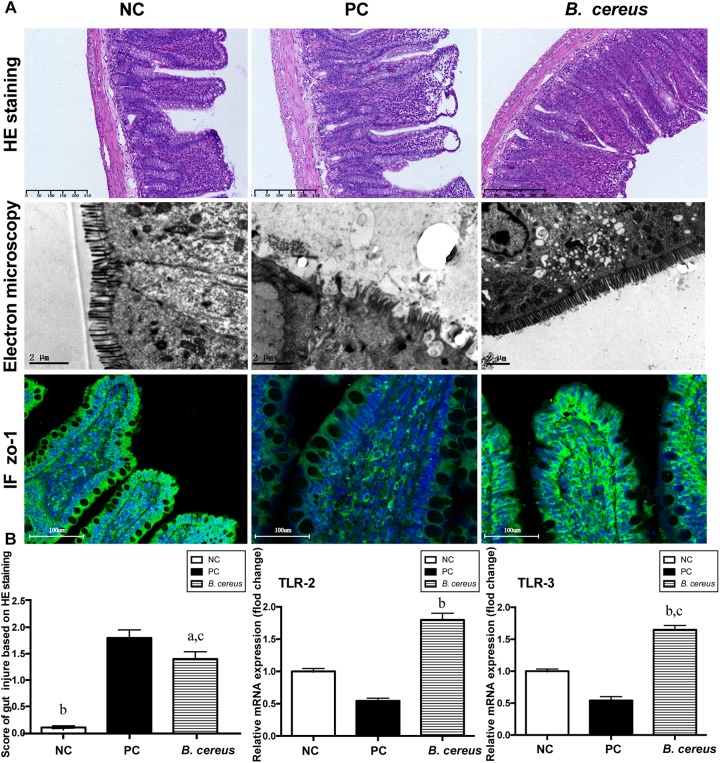
FIGURE 2.
Pretreatment with B. cereus reinforced the intestinal barrier function. (A) Representative images of H&E staining of the terminal ileum; ultrastructural and histological alterations in the ileum were assessed using ZO-1 immunofluorescence (IF) staining (×20). (B) Ileal inflammation was monitored based on the histology scores. Ileal mRNA expression of TLR-2 and TLR-3 was determined by quantitative PCR. All data are presented as the mean ± SEM. (NC group, n = 7; PC group, n = 6; B. cereus group, n = 7). aP < 0.05 and bP < 0.01 compared with the PC group, cP < 0.05 for the comparison of the B. cereus group with the NC group. NC, negative control; PC, positive control.

