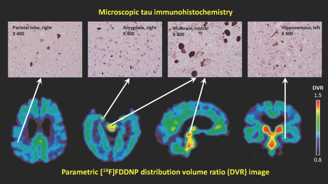
FIGURE 2.
Top panel: immunohistochemistry photomicrographs (×400) of parietal cortex, midbrain, amygdala, and hippocampus show the presence of tau neuropathological deposits in these regions. Bottom panel: representative transaxial (2 sections), sagittal (middle), and coronal sections (right) of [F-18]FDDNP-PET images with high signals in the periventricular subcortical regions, amygdala, and midbrain. Warmer colors (red and yellow) show areas with higher [F-18]FDDNP binding signals.

