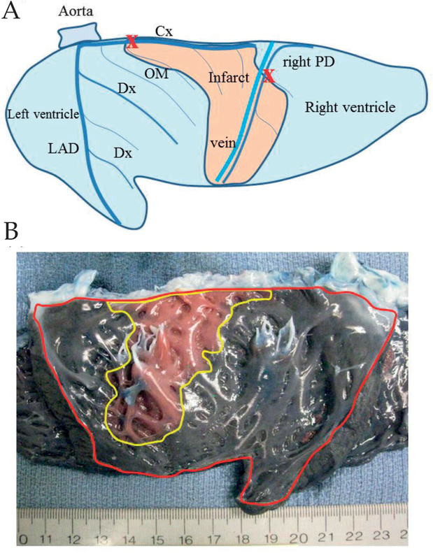
Figure 1:
A) Schematic epicardial view of the left and right ventricle after sectioning through the ventricular septum. A posterolateral myocardial infarction (flesh-colored area) was created by ligating both the main circumflex coronary artery (Cx) just distal of the first obtuse marginal artery (OM) (marked X) and the right posterior descending artery (right PD) at the level of 1.5 cm distal from the atrioventricular groove (marked X). Dx: Diagonal branch;LAD: Left anterior descending artery. B) Photographic endocardial view of the left and right ventricle after sectioning through the ventricular septum (left ventricle outlined in red) in an acute group animal after creating a myocardial infarction. Infarct area (outlined in yellow) is clearly delineated by the injection of Evans blue dye.

