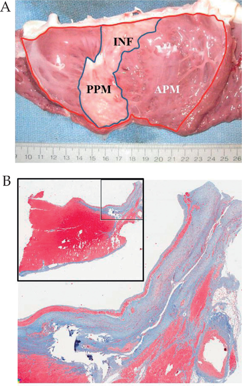
Figure 2:
A) Gross anatomic view of the endocardial side of the opened left ventricle, demonstrating the location of the myocardial infarction, which involves the entire posterior papillary muscle and extends up to the mitral annulus. Infarct size was determined by the ratio of the infarct area (INF; blue line) and the area of the entire left ventricle (red line). B) Masson’s trichrome staining demonstrating the transmural nature of the infarct. APM: Anterior papillary muscle; PPM: Posterior papillary muscle.

