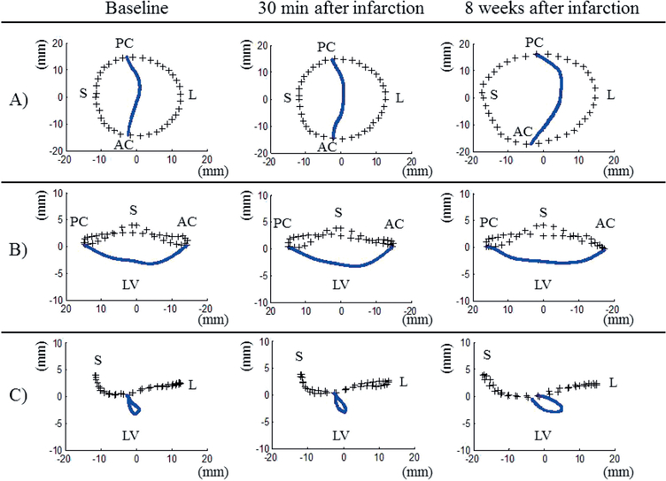
Figure 4:
Average three-dimensional plots of mitral valve annual (+++) and leaflet coaptation (blue line) that resulted from the mitral valve segmentation algorithm described in the text. A) Valve viewed from above. B) Septolateral view (looking over the saddle horn towards the posterior annulus). C) Intercommissural view. Plots were created at the time of baseline, at 30 min after infarction, and at eight weeks after infarction. Note the progressive annular dilatation, annular flattening and posterior displacement of the coaptation line over time. AC: Anterolateral commissure; PC: Posterolateral commissure; S: Septum (middle of anterior area); L: Lateral (middle of posterior area); LV: Left ventricle.

