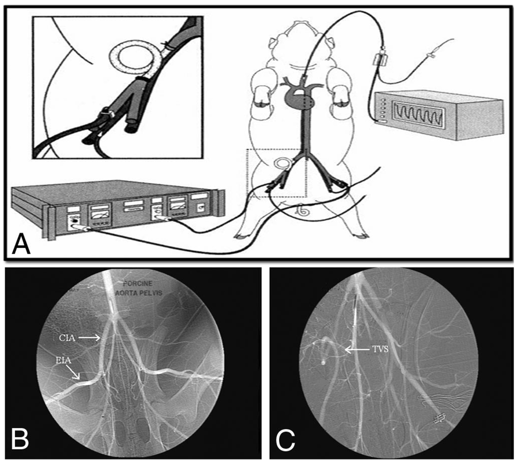
Figure 1.
(A–C)Experimental animal set up (A). The right external iliac artery is exposed and ligated. After a predetermined ischemic time, a temporary vascular shunt (TVS) is placed to reestablish flow (inset). Continuous ultrasonic flow confirms shunt patency and continued reperfusion. Arteriography shown here demonstrates pre- and post-TVS placement. Normal anatomy is shown with common iliac (CIA) and external iliac (EIA) arteries identified (B). After the predetermined ischemic time, a TVS (arrow) was placed in the artery and distal perfusion reestablished (C). (Reprinted with permission from J Trauma. 1999;47:64–71).

