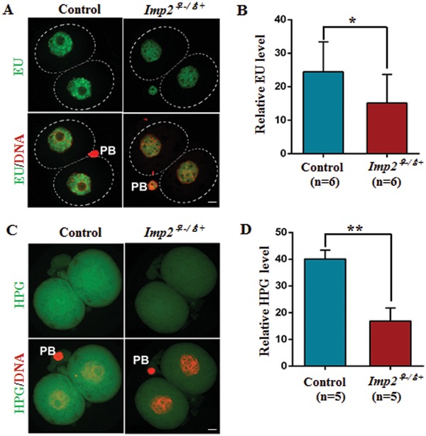
Figure 6.

Deletion of IMP2 disrupts the transcriptional and translational activity in 2‐cell‐stage embryos. A) Confocal image showing newly synthesized RNA by EU staining in control and 2‐cell‐stage Imp2−/− female embryos. Scale bar, 20 µm. B) Quantification of the fluorescence in newly synthesized RNA in control and Imp2−/− female 2‐cell‐stage embryos by EU incorporation. More than 10 embryos were observed for each genotype with six replicates. n = 6 mice for each genotype. Error bars indicate the SEM. *p < 0.05, Student's t‐test. C) Confocal image indicating the protein synthesis in control and Imp2−/− female 2‐cell‐stage embryos incorporating HPG. Scale bar, 20 µm. D) Quantification of the fluorescence in nascent protein synthesis by HPG incorporation in control and Imp2−/− female 2‐cell‐stage embryos. More than 10 embryos were observed for each genotype with six replicates. n = 5 mice for each genotype. Error bars indicate the SEM. **p < 0.01, Student's t‐test.
