
Figure 11.
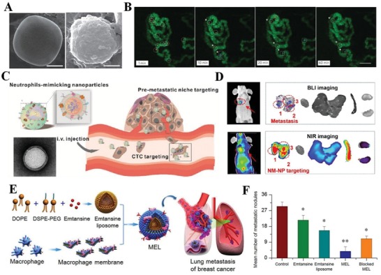
Cell membrane‐based nanotherapeutics for the inflammatory tumor environment. A) Representative SEM images of a bare NPS surface (left) and an NPS camouflaged with leukocyte‐derived membranes (right). Scale bars, 1 µm. B) Representative images of circulating and adherent LLVs (red, circles) and NPS (blue, squares) in the melanoma microvasculature (green) at different times (scale bar 100 µm). Reproduced with permission.[qv: 30c] Copyright 2013, Nature Publishing Group. C) Schematic representation of carfilzomib‐loaded NM‐NPs (NM‐NP‐CFZ) designed for neutralizing CTCs in circulation and preventing early metastasis. D) Dual‐mode imaging of NM‐NP‐CFZ‐treated mice with obvious metastasis 14 d after luc+ 4T1 cell injection in vivo and ex vivo. Reproduced with permission.72 Copyright 2017, American Chemical Society. E) Scheme of macrophage membrane‐coated emtansine liposomes (MELs) with specific targeting to suppress lung metastasis in breast cancer. F) The average number of macroscopic lung metastatic nodules from each treatment group, *p < 0.05, **p < 0.01. Reproduced with permission.112 Copyright 2016, American Chemical Society.