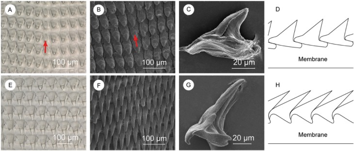
Fig 4. Description of central and lateral denticles.
(A and E) Optical microscope images of central, and lateral denticles. SEM images of (B) central denticles, (F) lateral denticles, (C) a single central denticle still showing some fibrils on the base, and (G) a clean single lateral denticle. Schematic representation of the (D) central, and (H) lateral denticles’ packing. Red arrows indicate the rachidian tooth where visible.

