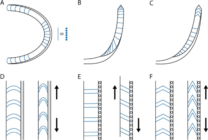
Fig 9.
Schematic representation of the odontophore sections: (A) transverse section, (B) coronal section, and (C) sagittal section. Schematic representation of the elastic behavior on different position of the odontophore, the forces inducing the deformation are reported as arrows: (D) transversal posterior region, (E) ventral sagittal region, and (F) dorsal sagittal region.

