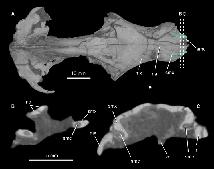
Fig 6. Pseudotherium argentinus, the septomaxillary canal.
(A) 3D volumetric rendering of dorsal view of skull showing septomaxillae in aqua tint (A), and dashed lines that indicate positions of cross sectional CT image slices (B) and (C). Abbreviations: ir, broken incisor root; mx, maxilla; na, nasal; smc, septomaxillary canal; smx, septomaxilla, vo, vomer.

