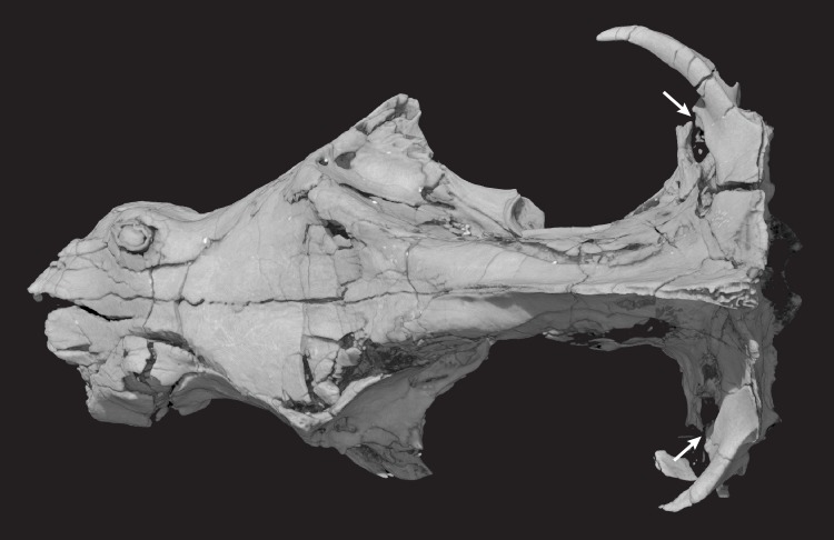
Fig 19. Pseudotherium argentinus skull in posterodorsal view illustrating open pterygoparoccipital foramina (indicated by arrows).
Each pterygoparoccipital foramen is almost entirely enclosed by the lateral flange of the petrosal (periotic) anteriorly and the squamosal posteriorly. The lateral flange and the squamosal do not contact, so that pterygoparoccipital foramen is laterally open. Because each foramen is open to a similar extent, this is not likely to be an artifact of post-mortem deformation.

